


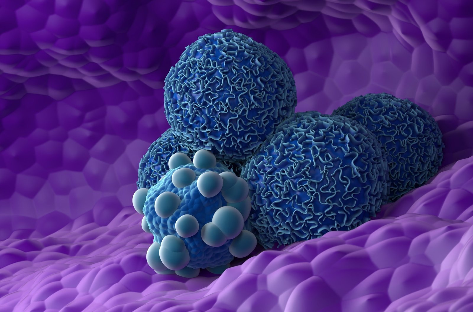
A new spatial map of pancreatic precancerous cells reveals striking discontinuities. Although PanIN cells may appear increasingly cancerous, the surrounding tissue may lack the stromal and immune changes necessary to support malignant transformation.
Study: Asynchronous evolution of epithelium and stroma distinguishes precursor lesions from pancreatic cancer. Image credit: Nemes Laszlo / Shutterstock
In a recent study published in the journal cancer discoveryresearchers found that the epithelial cells of pancreatic intraepithelial neoplasia (PanIN) gradually resemble cancer cells. However, the surrounding tissue environment was significantly different until the late stages of transformation. Therefore, the lack of complete stromal reprogramming may help explain why most PanIN lesions do not progress to pancreatic cancer.
Pancreatic ductal adenocarcinoma (PDAC) remains a deadly condition worldwide because the early disease goes undetected and is often diagnosed at a late stage, when cancer cells may have already begun to spread.
Scientists have focused on PanIN, the tiny precursor lesions that cause most pancreatic cancers. However, these lesions are difficult to study in humans because they require invasive procedures and are rarely obtained from healthy individuals. For this reason, researchers primarily rely on samples obtained from tumor-adjacent regions or genetically engineered mouse models with different molecular and anatomical features.
About research
In this study, researchers analyzed healthy donor pancreas obtained through a collaboration with Gift of Life Michigan. A broader donor program has yielded more than 150 pancreases from individuals aged 20 to 70. They also obtained pancreatic cancer samples from the University of Michigan. Advanced spatial transcriptomics and single-cell RNA-seq (scRNA-seq) have mapped the cellular and molecular changes underlying disease progression.
The researchers used advanced spatial genetic mapping techniques to examine 11 PanIN-containing donor samples, seven PDAC samples, and two adjacent normal pancreatic samples taken from cancer patients. A board-certified gastrointestinal pathologist examined all samples. They improved resolution by combining spatial transcriptomics with a comprehensive scRNA-seq dataset consisting of more than 200,000 cells from healthy, adjacent normal, and pancreatic cancer tissues.
The researchers used computer tools to identify epithelial cells, immune cells, and fibroblasts within the tissue. These tools performed tasks such as spot deconvolution, spatially informed clustering, pseudotime analysis, and ligand-receptor interaction analysis to analyze the samples.
The research team also studied protein expression using immunofluorescence. They analyzed publicly available Xenium and CosMx datasets to confirm their results. The research team also developed a three-dimensional organoid fibroblast minimodel in the lab using patient-derived pancreatic cancer organoids and fibroblast cells. These models simulated interactions between tumor cells and surrounding stromal cells.
Next, we assessed macrophage responses to tumor-derived signaling molecules. These approaches have allowed researchers to compare cells and their surrounding environment in healthy tissue, precancerous lesions, and invasive pancreatic cancer.
result
Epithelial cells within PanIN lesions showed molecular similarities to pancreatic cancer despite remaining non-invasive. Spatial transcriptome analysis revealed that PanIN epithelial cells exhibit gradual progressive changes that resemble those of pancreatic cancer. Notably, as the disease progressed, these cells exhibited increased RAS/MAPK or KRAS pathway activity, inflammatory pathways, hypoxia, and certain metabolic changes such as decreased oxidative phosphorylation and fatty acid metabolism.
Markers indicative of aggressive disease were also elevated in poorly differentiated tissues. These include Janus kinase (JAK) and signal transducer and activator of transcription 3 (STAT3) signaling molecules. Oncogenes such as keratin 17 (KRT17) and Wnt family member 7a (WNT7A) were also elevated.
Although epithelial cells exhibited tumor-like changes, the microenvironment around PanIN lesions behaved more like healthy pancreatic tissue than a tumor. The area adjacent to the PanIN lesion closely resembled the normal pancreatic duct. The area surrounding the PanIN lesions was full of plasma cells, immune cells that produce antibodies. Additionally, macrophages, which can promote inflammation and tumor growth, did not aggregate near PanIN lesions and remained distant from the epithelium.
These findings suggest that there is a potentially protective and regulated immune environment surrounding PanIN lesions, which may act as an important barrier to progression to cancer. Tumor cells showed the opposite pattern, with macrophages clustered near cancer cells and plasma cells dispersed throughout the tissue.
The researchers found that fibroblast populations enriched in tumors express high amounts of specific proteins. Examples include smooth muscle actin (SMA) and leucine-rich repeat-containing 15 (LRRC15). Such fibroblasts were associated with poor prognosis.
Laboratory-generated organoids demonstrated that cancer cells induce these fibroblasts to switch on lymphoid enhancer-binding factor 1 (LEF1). LEF1 is a gene involved in the wingless-associated integration site (Wnt) signaling pathway, which is associated with cancer growth and tissue remodeling. These effects were associated with cancer-associated molecular changes and transition to a more aggressive basal tumor-like phenotype.
The researchers also found that more than 60% of healthy donor pancreases contained PanIN lesions. This suggests that precursor lesions are surprisingly common but rarely progress to cancer.
conclusion
This finding suggests that PanIN lesions, although exhibiting tumor-like changes, have not yet developed the cancer-promoting microenvironment seen in pancreatic tumors. This may help explain why most PanIN lesions do not progress to cancer.
Future studies will require more advanced techniques to study pancreatic tissue at single-cell resolution. This will help us understand how molecular pathways such as WNT signaling affect cells surrounding PanIN epithelial lesions. Such efforts could improve our understanding of the natural barriers that can inhibit the progression of pancreatic precursor lesions to cancer, which could be targeted to inform cancer prevention strategies and reduce the cancer burden.
Click here to download your PDF copy.
