


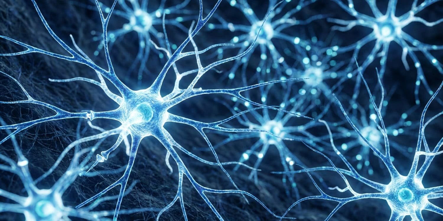
Recent research published in journals neuron provides evidence that a specific group of adrenaline-producing brain cells acts as a key driver of fear and anxiety behaviors. Activation of these cells causes immediate and long-term anxiety, and decreased activation of these cells reduces stress responses. These findings suggest that targeting these specific neurons may provide a more precise way to treat anxiety disorders in the future.
Anxiety disorders affect more than 300 million people worldwide and have a significant impact on their daily lives. Current treatments, including various medications and therapies, can cause side effects such as insomnia, irritability, and cognitive impairment. Relapses often occur when patients stop taking their prescribed medications. This ongoing challenge highlights the need for more specific and localized therapeutic targets within the brain.
Physical symptoms of stress, such as increased heart rate and rapid breathing, are regulated by regions in the lower brainstem. Within this region, the two neuronal populations are located very close together and share similar genetic profiles. A1 cells produce norepinephrine and C1 cells produce epinephrine, a chemical messenger also known as adrenaline.
These two cell types are highly intermixed, and previous scientific tools lacked the specificity to study them separately. Previous research has typically grouped them together and conducted research based on the assumption that they primarily control physical functions such as blood pressure rather than complex emotions. A research team based at St. Jude Children’s Research Hospital and led by Lindsay A. Schwartz with Carlos Fernandez-Peña sought to isolate the precise behavioral functions of these individual cell groups. They wanted to know whether C1 neurons directly modulate psychological anxiety, rather than just a physical stress response.
To separate C1 cells from A1 cells, scientists developed a special strain of genetically modified mice. They combined these mice with an engineered viral vector to create a biological logic gate that allowed them to selectively target only C1 neurons. The researchers used adult male and female mice between 6 and 20 weeks old for their behavioral experiments.
In the first test, animals were exposed to 30 minutes of physical restraint stress. They then examined RNA markers in brain tissue and found that this stressful event preferentially activated C1 neurons over A1 neurons. RNA is a molecule that helps carry out genetic instructions, and its presence indicates that the cell is recently active.
The authors then used optogenetics, a technique that uses focused light to control genetically modified cells. They implanted small optical fibers into the brains of mice to shine light directly on C1 neurons. During a 5-minute test in a 40 x 40 x 20 cm open field arena, light stimulation caused mice to spend significantly more time hiding in a corner.
The researchers also tested the mice in an elevated zero maze, a circular platform 100 centimeters in diameter that is 61 centimeters above the ground. The maze features alternating open and closed sections. Mice are naturally afraid of exposed areas and prefer walled areas.
When the researchers activated C1 neurons with light, the mice spent significantly less time outdoors, indicating an increased state of anxiety. Interestingly, when the researchers stimulated both A1 and C1 neurons simultaneously in another test, the mice stopped moving completely. This temporary behavioral arrest was not observed when targeting only C1 cells.
This difference provides evidence that the two adjacent cell types perform very different functions. The scientists then traced the physical connections of the C1 neurons to see where the signals were being sent. They found dense connections toward the ventrolateral periaqueductal gray matter. This midbrain region is well known for processing threats and triggering defensive behaviors such as freezing in place.
To test this particular pathway, the researchers moved an optical fiber to shine light only on the C1 nerve endings inside the midbrain. Activating this isolated connection produced a severe anxiety response in the elevated zero maze. The authors also found that short-term activation of this pathway caused persistent anxiety.
They stimulated C1 connections while the mice were in their home cages and then left completely undisturbed for 7 days. When tested in the elevated zero maze a full week later, these mice still showed elevated anxiety behavior compared to the control group. The researchers also tested a real-time place preference model in which mice avoided certain sides of the room when triggered by a light stimulus, and showed that animals found C1 activation highly unpleasant.
To observe natural brain activity, the researchers used fiber photometry, a method that measures calcium fluctuations to track cell firing in real time. As the mice explored the elevated zero maze, activity in C1 neurons increased rapidly at the exact moment the animals stepped into the scary open space. This natural brainstem activity directly excites downstream neurons in the midbrain.
Midbrain neurons remained active for several seconds after the mice entered the open zone. This persistent firing suggests that the circuit is constantly calculating environmental threats. To see if blocking this circuit could prevent anxiety, scientists used designer drugs to temporarily turn off C1 neurons.
They administered a special compound called DCZ at a dose of 500 micrograms per kilogram of body weight 15 minutes before the test. In one experiment, scientists exposed mice to an looming fear test in which a dark, expanding circle was projected from above to mimic a flying predator. Usually, rats freeze in fear at this sight. Inhibiting C1 neurons significantly reduced both the number of freezing events and their total duration.
The researchers also tested a fear conditioning model that combined a specific sound with a one-second mild electric shock to the foot. A few days later, the mouse froze when I played just that sound. Mice with suppressed C1 activity showed that this learned freezing behavior diminished much faster over time.
For the final behavioral test, the scientists combined a 30-minute period of physical restraint with an elevated zero maze. Typically, when pre-restrained, mice are almost completely hidden in the closed section of the maze. Turning off C1 neurons just before the restraint period completely abolished the stress-induced anxiety on this maze.
Using a modified rabies virus as a biological tracker, the researchers mapped all the upstream regions of the brain that send commands to C1 cells. They found that C1 neurons receive input from areas that regulate pain, breathing, sleep, and basic internal body conditions. This network arrangement tends to position C1 cells as central hubs that collect physiological data to inform emotional responses.
A potential limitation of this study is the use of only animal models. The human brain is much more complex, and human anxiety is influenced not only by innate predator avoidance but also by learned psychological factors. Functional connections between the brainstem and midbrain also exist in humans, meaning this circuit likely plays a similar role in humans, but human studies are needed to confirm this.
Another limitation is that the researchers pooled male and female mice together for the analysis. They did not independently analyze the data to look for differences by gender. Future research should investigate whether the operation of this anxiety circuit differs depending on biological sex.
The short lifetime of designer drugs used to inhibit cells also limited the scope of the study. When the researchers tried adding a delay period between the stressful event and the behavioral test, the anxiety returned as the medication wore off. Longer-lasting inhibition methods are needed to fully understand the long-term benefits of turning off these neurons.
Future studies should investigate how C1 neurons behave in different internal states. In one test with mice fasted in a new environment, stimulating C1 cells actually made the mice eat faster. This unexpected result suggests that this circuit may adapt its function based on extreme hunger or other pressing biological needs. The researchers hope to map exactly how interoceptive signals, such as changes in breathing and heart rate, enter C1 neurons and trigger panic.
The study, “Autonomic C1 neurons promote anxiety via vlPAG activation,” was authored by Carlos Fernández-Peña, Rachel L. Pace, Lourds M. Fernando, Heather Sheppard, Brittany G. Pittman, and Lindsay A. Schwarz.
