


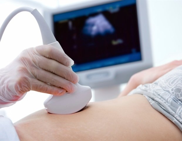
Current fetal monitoring tools are inadequate for doctors to closely track high-risk pregnancies, so researchers at Stanford School of Medicine, the University of California, San Diego, and the University of Oxford have developed a wearable ultrasound patch to monitor such pregnancies. The patch is applied to the patient’s abdomen and provides continuous, real-time information about blood flow to the fetus and umbilical cord.
The device’s design and initial validation with dozens of pregnant patients was published on May 26. nature biotechnologyis expected to help doctors monitor conditions such as intrauterine growth restriction, which affects 10% of all pregnancies. This pregnancy complication results from insufficient blood flow through the umbilical cord, which limits oxygen and nutrients and slows fetal growth. In severe cases, doctors may deliver the baby early to allow for better development or to avoid stillbirth.
“There is nothing similar to our device on the market or in the scientific literature,” said lead study author Dr. Sheng Xu, professor of anesthesiology, perioperative and pain medicine at Stanford University. Xu previously served as a professor at the University of California, San Diego, where much of his research was conducted.
The study’s lead author is Dr. Geonho (Tom) Park, currently a postdoctoral fellow in anesthesiology, perioperative and pain medicine at Stanford University, but at the time the study was conducted he was a graduate student at the University of California, San Diego. So are Yizhou Bian, Hao Huang and Sai Zhou, graduate students at the University of California, San Diego.
Xu and Park continue to develop the ultrasound patch at Stanford University.
The ultrasound patch is a palm-sized, flexible adhesive sticker that is placed on the abdomen. It is connected by a cable to a computer that interprets the ultrasound data. Researchers believe the patch will initially be used on pregnant women in the hospital, but they hope to eventually develop a wireless version that allows doctors to monitor patients at home.
“Umbilical artery blood flow is one of the factors we look at closely when concerned about the health of the fetus in cases of placental insufficiency,” said Jane Chue, MD, a high-risk obstetrician at Stanford School of Medicine who will collaborate with Professor Xu’s team in further validation of the technique. But data is currently complicated to obtain, she added.
Need for fetal data
Unlike newer ultrasound patches, current diagnostic tools typically provide a short snapshot of the fetus’s condition.
Measuring blood flow with existing Doppler ultrasound machines not only captures data over a short time frame, but also requires a trained ultrasound technician and an appointment, complicating matters even for hospitalized patients, Chueh said.
Another current option for assessing fetal health is electrocardiography, a combined measurement of fetal heart rate and uterine contractions, data obtained from a monitor strapped to the abdomen. Chue’s team is introducing cardiac testing to women with high-risk pregnancies at Lucile Packard Children’s Hospital in Stanford.
“It’s really difficult to keep using it continuously,” Chueh said, noting that the device could give a false signal or no signal when the fetus is moving a lot. “Even for hospitalized patients, getting accurate readings three times a day can be labor-intensive.”
The monitor may show a decrease in the fetal heart rate due to actual changes or simply because the fetus is moving around, requiring regular checking and repositioning of the monitor. “This is extremely stressful for both patients and medical staff,” Chueh said.
More sophisticated monitoring equipment could help doctors better navigate the tightrope of deciding when to deliver a baby in a risky pregnancy. Giving birth early increases the risk of complications in a premature baby. But waiting to give birth can be even worse if the fetus isn’t receiving enough blood flow.
technical challenges
In developing the patch, researchers had to overcome many obstacles. Unlike most wearable devices, which measure information at or near the body’s surface, this ultrasound patch must collect and interpret information from deep within the uterus.
Also, everything they wanted to visualize is in motion. Not only does the pregnant patient’s body move, but the fetus practices inversions and the umbilical cord floats freely in the amniotic fluid. In a traditional Doppler ultrasound setup, the technician can change the position of the machine’s transducer to get a better view.
The researchers used a combination of strategy and innovation to tackle the problem.
We asked ourselves, “What would happen if we used an ultrasound device to irradiate the placenta in the area where the umbilical cord is attached?” Even though everything is moving, there is some stability in the umbilical cord of the place. ”
Geonho (Tom) Park, PhD, Postdoctoral Fellow in Anesthesiology, Perioperative and Pain Medicine, Stanford Medicine
The research team developed an image segmentation algorithm that can track in real time the end of the umbilical cord, which is secured to the placenta, a key element of the design.
After developing a prototype and testing that it worked as expected on a simulated mannequin, the researchers confirmed that it did not impart excessive acoustic or mechanical energy to the fetus. This device meets safety standards set by the US Food and Drug Administration, the American Institute of Ultrasound in Medicine, and the British Medical Ultrasound Association.
The next step was to verify the device. The researchers tested the patch on 62 pregnant women and compared the results with those from a standard Doppler ultrasound machine. The new patch and the old machine gave statistically equivalent results.
This patch can image all three major blood vessels in the umbilical cord (two arteries and one vein). It also measures blood flow through the fetus’s major arteries and measures fetal anatomy such as head circumference, abdominal circumference, and femur length to help estimate fetal weight, which is a key indicator for diagnosing growth problems. The image segmentation algorithm accurately tracked the umbilical cord without changing its position on the mother’s abdomen, even as the mother moved around. It also worked regardless of the location of the placenta within the uterus.
Detection of serious complications
While testing the device, the research team discovered something unexpected in one study participant.
“She was in her 28th week of pregnancy, which is still quite early, and initial tests showed that the fetal heart rate was normal. Later, we found that the blood flow signals were quite abnormal,” Park said. “I thought, ‘Maybe there’s something wrong with the device,’ so I checked everything, and everything seemed fine. I showed the data to the doctors who were there, and they agreed that the fetus might be at risk.”
The participants’ ultrasound data showed large fluctuations in blood flow through the umbilical cord, in contrast to the steady blood flow of healthy patients at a similar stage of pregnancy.
Follow-up testing confirmed that the participant had severe placental insufficiency. She gave birth four days later by caesarean section under close supervision of doctors. The baby went to the neonatal intensive care unit and made an uneventful recovery.
next step
The research team plans to further refine and validate the use of the ultrasound patch at Stanford Medicine. In addition to building a wireless version of the device, they plan to test it more extensively on patients with other pregnancy complications that involve poor blood flow, such as congenital heart disease and chronic hypertension.
“Right now, for patients with these high-risk pregnancies, it can be difficult for doctors to get the information they need when they need it,” Chueh said. “I think this device will make it easier to get that information.”
“We’ll start with inpatients, but perhaps one day we’ll be able to use it in outpatients as well,” she added.
This research was funded by grants from the National Institutes of Health (grants 1R01EB033464-01 and 1R01HL171652-01) and grants from Wellcome Leap and Accelerated Innovation to Market at the University of California, San Diego.
sauce:
Reference magazines:
Park, G. others. (2026). Fetal monitoring of high-risk pregnancies using a wearable ultrasound patch. nature biotechnology. DOI: 10.1038/s41587-026-03140-1. https://www.nature.com/articles/s41587-026-03140-1
