


New research published in journal neuroimage It suggests that certain mathematical measures applied to brain scans can successfully track the brain’s ability to quiet its own activity. By observing how alcohol consumption changes brain dynamics in both rodents and humans, scientists found evidence that this indicator is significantly reduced when the brain’s inhibitory system is activated. These discoveries provide new non-invasive tools to help doctors and researchers monitor brain function in living subjects.
A team of scientists from the University of Pennsylvania, the University of North Carolina, and research institutions in Germany collaborated on the project. Their goal was to better understand how to track brain activity patterns in different species.
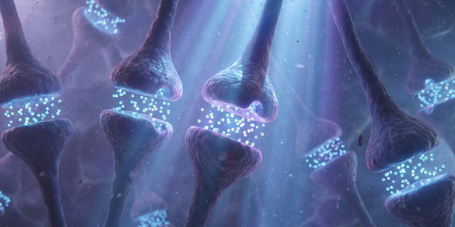
The brain functions through a delicate balance of excitatory signals that increase activity and inhibitory signals that act like a braking system. Neural inhibition is an important braking process for this, helping to stabilize brain networks and filter out excess noise. Disturbances in this system tend to be associated with a variety of developmental disorders and mental health conditions, including depression, autism, and schizophrenia.
It is very difficult to directly measure neural inhibition in the living human brain because traditional methods can only examine small parts of the brain at a time. For this reason, researchers have been looking for indirect markers seen in standard brain scans of the whole brain. One of the proposed markers is the Hurst index, which is a mathematical calculation applied to data from functional magnetic resonance imaging. Functional magnetic resonance imaging (fMRI) is a common technique that measures brain activity by detecting changes in blood flow over time.
The Hurst index focuses on the long-term temporal correlation of brain signals. In other words, it measures how predictable and structured brain activity is from one moment to the next. A higher Hurst index suggests a more stable and controlled brain network, which scientists believe reflects strong neural inhibition. Lower values indicate more irregular and noisy brain activity. So far, the evidence linking this calculation to actual suppression is mostly correlational.
The research team wanted to test whether this mathematical marker actually changes when directly manipulating the brain’s inhibition levels. They decided to use alcohol as a tool to change brain chemistry, since alcohol is widely known to depress central nervous system activity. Alcohol accomplishes this primarily by interacting with specific docking stations on brain cells known as GABAA receptors. These receptors respond to a chemical called GABA, which is the main inhibitory messenger in the brain.
To thoroughly test this idea, the authors began by examining animal models to minimize the effects of different environments and genetics. They analyzed brain scan data from a total of 38 laboratory rats bred for this specific purpose. Three rats were excluded from the final analysis because they moved too much during the scanning process, resulting in a final sample of 35 rats. These animals were young adults, 45 or 80 days old.
During the experiment, rats were safely anesthetized and placed in an animal-sized fMRI scanner. The researchers recorded continuous resting-state brain activity divided into 15-minute blocks over a period of 75 minutes. The scientists first injected the rats with simple saline to establish a baseline control. After this, they administered three progressively larger doses of ethanol, the type of alcohol found in common drinks.
After calculating the Hurst index for different brain regions, the scientists observed significant changes. After injections of larger amounts of alcohol, particularly 2 and 4 grams per kilogram, the Hurst index was significantly reduced throughout the brain compared to the initial saline injection. This decline indicates that brain signals became less structured and more irregular as alcohol strengthened neural inhibition.
This effect was particularly pronounced in the sensory and emotional centers of the rat brain, including the auditory, visual, and entorhinal regions. Subcortical regions deep in the brain, such as the cerebellum and amygdala, also showed significant reductions in the Hurst index. The researchers then compared these brain map changes to known maps of GABAA receptors in rat brains. They found a strong negative correlation. This means that the brain regions with the highest density of these inhibitory receptors experienced the greatest reduction in the Hurst index.
To see if these effects also occur in humans, the authors conducted a human study using a large repeated measures design. The sample included 11 healthy adult volunteers aged 24 to 33 years. These participants were scheduled to attend 10 separate testing sessions over several weeks. Five of these visits involved the consumption of alcohol and five did not involve the consumption of alcohol. The order was randomized for each person.
During the alcohol session, participants drank a mixture of vodka and orange juice on an empty stomach. Beverage volume was tailored to each person based on height, weight, and gender to reach a target blood alcohol content of 0.08%. After drinking the drink, volunteers lay inside a human fMRI scanner and focused on a simple white cross on a black screen. In the non-alcoholic session, exactly the same scanning procedure was followed without the use of intoxicating drinks.
Similar to animal studies, the researchers found that exposure to alcohol significantly lowered the average Hurst index across the human cerebral cortex. This provides evidence that markers respond to pharmacological changes consistently across different species. In humans, the most dramatic reductions occurred in association areas, outer regions of the brain involved in higher-order processing and complex thinking.
The research team also compared human brain scans with chemical maps obtained from positron emission tomography scans. These specific types of medical scans show exactly where different chemical receptors are located in a living human brain. The authors found that areas with high concentrations of GABAA receptors had the greatest decrease in Hurst index during alcohol sessions. Alcohol also interacted with receptors for other brain chemicals such as dopamine and serotonin, which showed a secondary correlation with changes in brain scans.
Several limitations should be kept in mind when interpreting these results. One of the major challenges when using the Hurst exponent is its sensitivity to physical movement during brain scans. Alcohol tends to make both humans and animals fidgety or move slightly, which can introduce errors in fMRI data.
The researchers applied rigorous mathematical corrections to account for this movement, and also excluded human scanning sessions in which participants moved too much. Even with these corrections, the effects observed in the human brain were relatively subtle when looking at individual, small brain regions. Alcohol is also known to alter heart rate and blood flow, which may indirectly alter fMRI signals.
Another limitation is that the receptor maps used for comparison were from separate groups of humans and rats, rather than the individuals actually scanned in this study. This means the researchers were unable to measure precise individual differences in receptor location. Future studies may use a combination of scanning techniques on the same individuals to separately understand how alcohol affects brain signals.
Future research could also investigate how changes in the Hurst index relate to actual changes in human behavior. The study focused strictly on physical brain measurements, rather than testing participants’ memory or self-control. Understanding how a decline in the Hurst index is related to the impulsivity and poor decision-making commonly seen after drinking alcohol would provide a more complete picture of neural inhibition.
The study, “Alcohol affects fMRI markers of neural inhibition in humans and rodents,” was authored by Monami Nishio, Xinyi Wang, Eli J. Kornblath, Songho Lee, Yen-Yu-Ian Shi, Nicola Palomero Gallagher, Michael J. Arcaro, David M. Lydon Staley, and Alison P. McKee.
