



Globally, humans currently produce a staggering 450 million tonnes of plastic each year. We use plastic in almost every area of our lives, from food and drink containers to cosmetic packaging, sewer pipes, window frames and polyester clothing. Almost a quarter of it is then released into the environment, where it breaks down very slowly into tiny pieces.
These microplastics (particles between 1 micrometer and 5 millimeters wide) have been found in the deepest parts of the oceans, on the tops of the highest mountains, in sparsely populated polar regions, and even inside the human body.
It is extremely difficult for living organisms, including humans, to avoid ingesting microplastics. Once these microplastics pass through the lining of the digestive tract and enter the bloodstream and other tissues, they remain in the body. Until now, it has been difficult for researchers to accurately assess whether this is happening.
Our research team has developed a new technique to localize microplastics within living organisms without dissecting them. We tested this in earthworms and found that microplastics between 5 and 53 micrometers in size easily passed through the lining of the intestine and did not enter other tissues of the earthworm.

Earthworms are essential to healthy soil and feed where microplastics accumulate.
(Unsplash/Shippacorn/Yamkashicorn)
pollution problem
The question of whether microplastics are excreted simply through the gastrointestinal tract or through the lining of the intestines has been a difficult question for researchers to answer. This is because the size of microplastic particles must be less than 83 micrometers. This is 10 times smaller than the head of a sewing pin.
Researchers are being forced to dissect the organisms’ tissues to determine whether such tiny particles could have passed through the lining of the digestive tract and entered other tissues. Because microplastics are ubiquitous, it is very difficult to prevent dissected samples from becoming contaminated with more microplastics, and it is difficult to accurately measure what was originally present.
Read more: How to turn plastic waste into vinegar: A solar breakthrough
To avoid contaminating tissue samples, our research team collaborated with the Canadian Light Source in Saskatoon to take very high-resolution X-rays on the earthworms using a technique called synchrotron-based microcomputed tomography.
bright white particles
We chose to work with earthworms because they are essential to healthy soil, constantly moving and eating soil where microplastics can accumulate. This makes it a useful species to study how soil organisms interact with microplastic pollution.
We fed earthworms soil containing particulates ranging in size from 5 to 22 micrometers, or from 45 to 53 micrometers. These microplastics are coated with barium salts, meaning they appear as bright white particles inside the earthworm’s body under X-rays.
In this three-dimensional image of an earthworm, microplastics appear as bright particles against a background of dark tissue.
Traditional X-rays do not provide the detail needed to locate microplastics within the earthworm’s body. Microcomputed tomography allowed them to create highly detailed three-dimensional images of earthworms. In this image, microplastics are visible as bright particles against the background of the earthworm’s dark tissue.
This allowed them to count the microplastics in the earthworms’ intestines and determine whether the microplastics ingested by the earthworms were transferred from the intestines to other tissues.
Microplastics do not exist outside the intestine
We observed a total of 2,779 microplastics in the digestive tract of C. elegans fed contaminated soil. No microplastics were found outside the digestive tracts of these insects.
This provides conclusive evidence that microplastics ranging in size from 5 to 53 micrometers do not easily cross the intestinal lining and invade other tissues of the worm.
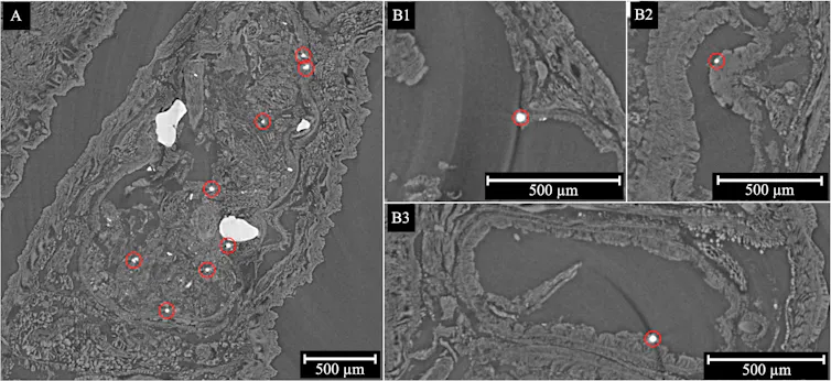
Synchrotron-based microcomputed tomography images of earthworms exposed to barium titanate glass microspheres. In panel A, red circles indicate individual microspheres present within excreta within the gastrointestinal tract. Panels B1 to B3 show microspheres in direct contact with the intestinal epithelium in areas where excreta are absent.
(Nick Lewin)CC BY-NC
This is also the first time synchrotron-based microcomputed tomography has been used to track the movement of microplastics within an organism.
human digestive tract
Are our findings applicable to humans and other species?
Although we should be cautious about extrapolating research on earthworms to other species, this study suggests that we may need to better evaluate the ability of our digestive tract to act as a barrier to indigestible food components.
The study also highlights the importance of developing techniques that can localize microplastics within living organisms without dissecting them. These techniques, such as synchrotron-based microcomputed tomography, provide more definitive results on how microplastics move within organisms.
This will help improve the risk assessment of microplastics for all life, including humans.
