


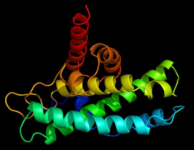
Structural biologists at the University of Cincinnati have become the first in the world to visualize a key cellular protein as part of a newly published study from the School of Medicine.
The Seegar Lab used cryo-electron microscopy at UC’s Advanced Structural Biology Laboratory to visualize for the first time the structure of the regulatory protein iRhom1 bound to the ADAM17 enzyme. This follows work from the lab published last year that visualized the structure of ADAM17 bound to iRhom2.
ADAM17 enzyme activity is essential for proper tissue development and immune responses in humans, and regulation of its activity has become a drug target in the treatment of chronic inflammatory diseases. Extracellular domain shedding is a fundamental biological process in which enzymes such as ADAM17 rapidly cleave and release other protein targets from the cell surface, altering cell-cell communication.
This latest study, published in Cell Reports, therefore identified structural elements in both iRhom1 and iRhom2 that function as molecular relays, transmitting information across the cell surface and linking intracellular signaling to activation of the ADAM17 enzyme on the cell surface.
ADAM17 is rapidly activated in response to changes in intracellular signaling networks, but how these signals are transmitted across the cell membrane to where ADAM17 resides remains a long-standing question in the field. ”
Dr. Tom Seeger, Corresponding Author, Assistant Professor, Department of Molecular and Cellular Biosciences, Distinguished Scholar at Ohio State
The study’s co-lead authors are Dr. Joe Maciag, a research associate in the Seeger Institute, and Joe Angbari, a third-year cancer and cell biology graduate student.
The Seegar Lab also revealed new insights into why iRhom1 and iRhom2 proteins are considered master regulators of ADAM17, which exists only in complex with iRhom1 and iRhom2. They found that the structures of iRhom1 and iRhom2 are identical, as well as their responses to intracellular signals, leading to a unified model of enzyme activation.
“Although their structures are strikingly similar, their functions are diverse. Their ability to maintain different roles despite their overall structural similarity is most likely due to subtle differences in their sequences that aid preferential recognition and cleavage of substrates,” Maciag said.
How they know which functions or tasks to perform and why they make different decisions is expected to be studied in more detail in the future. “That’s something that’s been missing in our field for 30 years,” Seger said.
Furthermore, iRhom proteins, particularly iRhom2, appear to be drivers of ADAM17 specificity and may therefore further serve as novel drug targets for treating chronic inflammatory diseases.
The researchers also looked at iRhom1 mutations identified in patients with cardiomyopathy.
They found that this variant was completely defective in supporting the iRhom1-ADAM17 function. “We found that the iRhom1 protein is likely not folded properly,” Ungvary said. “Because the structure of the protein is incorrect, its function is disabled.”
In this case, ADAM17 is unable to function properly or reach targets near the cell surface. Dysregulation of ADAM17 activity is thought to be involved in a wide range of diseases, including chronic inflammation, cancer, and neurodegenerative diseases.
“Notably, this phenotype is different from that observed in animal models and may more accurately reflect the consequences of iRhom1 dysfunction in humans,” Seegar said. “This is some of the first understanding of how this biology differs in humans and animal models.”
sauce:
Reference magazines:
Ungvallee, Genesee; others. (2026). Structural basis of ADAM17 activation by the iRhom1 pseudoprotease. cell report. DOI: 10.1016/j.celrep.2026.117309. https://www.cell.com/cell-reports/fulltext/S2211-1247(26)00387-6
