


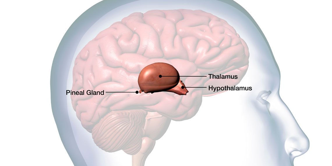
A new statistical study reveals that people diagnosed with a mental illness tend to have physically smaller pineal glands than people without a mental illness. This comprehensive study, published in Acta Psychiatrica Scandinavica, also shows that this structural difference does not seem to directly impact a person’s sleep quality.
The pineal gland is a small pea-shaped structure located deep in the brain. Its main function is the synthesis and release of melatonin. This hormone helps control your body’s internal clock, called the circadian rhythm. Circadian rhythms determine many physiological changes over the daily cycle and influence body temperature, metabolism, and immune function.
When the sun goes down and it becomes dark, the pineal gland increases melatonin production, signaling to the body that it’s time to rest. This disruption of the nighttime cycle is very common among people diagnosed with severe mental health conditions. Specifically, patients with major depressive disorder, bipolar disorder, and schizophrenia routinely experience extreme insomnia and altered sleep patterns.
The pineal gland is made up almost entirely of pineal cells, which are specialized cells responsible for producing melatonin. Because of this direct body structure, researchers believe that the overall size of the gland may determine a person’s total ability to produce sleep-promoting hormones. In theory, physically smaller glands house fewer hormone-producing cells.
Previous attempts to measure the pineal gland in psychiatric patients have yielded conflicting results. Some brain imaging studies have reported smaller volumes in patients with depression and schizophrenia, while others have found no differences in anatomical size.
Researchers Sophie Borwig and Christian H.R. Jensen from the Neurobiology Research Unit at Rigshospitalet in Copenhagen, Denmark, wanted to resolve these conflicting reports. They designed a meta-analysis. This is a research method that pools data from many separate studies into one mathematical model. This approach helps researchers discover broader patterns that may be hidden within the noise of isolated datasets.
The researchers systematically searched academic databases and found studies that used magnetic resonance imaging (commonly known as MRI) to measure gland size. They divided the research they collected into two different analyzes based on the specific biological questions they wanted to answer.
The first analysis focused on anatomical differences by comparing brain scans of people with formally diagnosed mental illnesses to scans of healthy individuals. The study group included more than 1,700 participants in total. The second analysis used data from more than 700 participants to examine the association between pineal gland size and specific sleep measures.
In an anatomical comparison, Bollwig and Jensen found that people with mental illness had an overall reduction in pineal gland volume. To understand whether this reduction was caused by the shrinkage of the glands or simply the presence of cysts, the research team took a closer look at the functional organization. A pineal cyst is a small pocket of fluid within the gland and is generally considered a harmless structural abnormality.
The researchers calculated the specific prevalence of these cysts and found no difference in frequency between psychiatric patients and healthy participants. When only noncystic tissue was examined, this functional mass was also found to be reduced. This means that the actual hormone-producing structures were physically smaller in the patient group.
The extent of this anatomical reduction varies widely depending on the specific psychiatric diagnosis. People with mood disorders such as major depressive disorder and bipolar disorder had slightly reduced pineal gland volume. In contrast, patients on the schizophrenia spectrum showed an approximately two-fold reduction.
To see if these physically small glands actually lead to sleep problems, the researchers turned to a second pool of data. These studies measured sleep variables using self-report questionnaires, wearable motion trackers, and overnight monitoring in specialized sleep laboratories.
The pooled data showed that the relationship between a larger pineal gland and sleep quality was weak and not statistically significant. There is wide variation in how different studies measure sleep quality, making it difficult to draw absolute conclusions. Still, current data suggest that the anatomical size of the gland does not directly influence a person’s daily sleep quality.
The researchers noted that these findings highlight the multifaceted nature of human sleep. A human’s ability to fall asleep and stay asleep depends on various nervous systems as well as psychological stressors, environmental factors, and melatonin production. Even if the physically smaller pineal gland produces slightly less melatonin, other networks in the brain may compensate to maintain basic sleep patterns.
The study also provides hints about the biological origins of certain mental health conditions. Some of the studies analyzed included people who were classified as being at high clinical risk of developing psychosis, but who had not yet developed the full disorder. These high-risk people already showed reduced pineal gland volume.
Because this structural difference appears before severe psychiatric symptoms appear, researchers have proposed that a smaller pineal gland may be a genetic trait of neurodevelopment. This idea is supported by recent genetic studies showing that genes that influence pineal gland volume overlap significantly with known genetic risk factors for schizophrenia. This is likely an inherent vulnerability rather than a by-product of long-term illness or use of psychiatric drugs.
Other biological mechanisms may also explain the volume variation. Medical professionals are increasingly recognizing that neuroinflammatory processes are involved in both mood disorders and schizophrenia. Chronic inflammation can affect the pineal gland through oxidative stress, but scientists have not yet determined whether this physiological process actively shrinks the tissue over time.
Although the exact mechanism remains unknown, the researchers acknowledged that the available evidence has some limitations. For example, the imaging studies included in the review were completely cross-sectional. This type of study takes a single snapshot at a single point in time, making it impossible to determine the long-term progression of anatomical changes in the brain.
Additionally, many of the original studies relied on older magnetic resonance imaging scanners with low spatial resolution. Medical professionals then had to manually trace the outline of the pineal gland on these old scans to calculate its volume. This manual process is subject to human error and measurement discrepancies between different research centers.
Going forward, scientists need longitudinal studies that track brain anatomy, sleep patterns, and hormone levels in the exact same individuals over many years. By observing people from childhood and adolescence to adulthood, scientists may be able to determine exactly when the pineal gland stops growing in people who later develop mental illness. Understanding this developmental timeline could ultimately allow physicians to identify biological risk factors long before a clinical crisis occurs.
The study, “Pineal gland volume, sleep quality, and psychiatric disorders: a systematic review and meta-analysis,” was authored by Sophie Bolwig and Kristian HR Jensen.
