


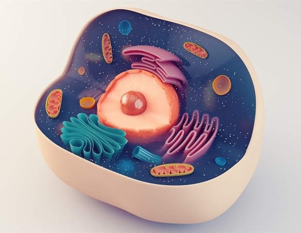
Cells are dense, dynamic places, with thousands of molecules interacting in tight spaces. Until now, scientists had no reliable way to watch many of these molecular interactions occur.
Researchers at the University of Illinois at Chicago have developed a new imaging method that allows researchers to see previously hidden enzyme activity in small areas throughout cells. The survey results are Proceedings of the National Academy of Sciencesopening new possibilities for understanding how cells process information.
This could help scientists better understand how drugs work or fail by showing exactly where cellular signals occur in real time.
We’ve discovered a cool lens into the characteristics of very small molecules used by cells. ”
Gary Mo, study co-author and UIC associate professor of pharmacology, regenerative medicine, and biomedical engineering
Other authors of the study are Kriti Srivastava, Kevin P. Schnur and Kay Petruzzi of UIC.
Why biosensor imaging sometimes fails
Scientists have long used tools called biosensors, fluorescent molecules that sense events inside cells and report on their activity.
“A biosensor is anything that can sense changes in the environment inside a cell,” says Srivastava, an assistant professor of medicine. If a cell is like a city, and each molecule is one of its inhabitants, then biosensors are like private investigators that track and report on people’s actions. They are the molecular eyes and ears that tell scientists what’s going on in living cells.
Biosensors can be positive or negative, meaning they “turn on” or “go dark” when they sense an event within a cell. Unfortunately, negative biosensors have often been unavailable to scientists.
As Mo explains, the problem is like wearing green clothes in front of a green screen: important details disappear. “These biosensors are dark, so some of the foreground where the action is is blended into the background,” Mo said. As a result, some regions with high enzyme activity can falsely appear identical to regions with no activity, he explained.
Turn unusable signals into clear images
To address this, the UIC team developed a methodology called FNICI (FINICI). This approach inverts each negative biosensor optical reading to a positive readable reading. This allows researchers to use existing negative biosensors without having to redesign them from scratch, which can take years.
The research team used FINICI to image the activity of three molecules: Src kinase, Syk kinase, and cGMP.
For Src kinase, a protein associated with cancer and cell motility, the researchers found bursts of activity in small regions of cell membranes that contain cholesterol-rich lipid rafts. Some of these active regions appear briefly before lysis, while others persist longer, a difference not seen in traditional whole-cell measurements.
The researchers also discovered that the signaling molecule cGMP forms small clusters that are quickly overwhelmed as the signal spreads inside the cell.
In immune cells, the enzyme Syk was most active near the scaffolds inside the cells, rather than near the receptors that activate it.
Taken together, these findings show that important signals are controlled by where molecules act within the cell. As with real estate, location is very important. “I have to be in the room to do my job,” Mo said. “If the enzyme isn’t in the right place, it doesn’t matter whether the enzyme is active or not. It works, but it doesn’t really work.”
Moe and Srivastava said the implications of their discovery extend far beyond the lab. Many drugs are designed to target enzymes or signal transduction pathways, and their effectiveness may depend on this location issue.
“Cell signaling determines how drugs work. Drug molecules interact directly with molecules within the cell. Visualizing these details is an important step in helping us understand and improve how drugs work,” said Mo.
sauce:
University of Illinois at Chicago
Reference magazines:
Srivastava, K. others. (2026). We directly employ inverse biosensors to image the enzymatic activity of living cells within nanodomains. Proceedings of the National Academy of Sciences. DOI: 10.1073/pnas.2531118123. https://www.pnas.org/doi/10.1073/pnas.2531118123
