


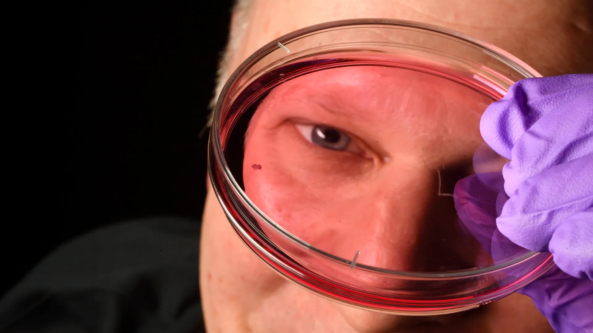
Scientists at Johns Hopkins University have uncovered how humans develop sharp central vision before birth, identifying carefully timed interactions between vitamin A-derived molecules and thyroid hormones in the retina. The discovery challenges decades-old explanations of how important light-sensing cells are formed and could guide future treatments for macular degeneration, glaucoma and other vision-damaging diseases.
The study, which was based on retinal tissue grown in the lab, Proceedings of the National Academy of Sciences.
Lab-grown retina reveals how clear vision is formed
“This is an important step toward understanding the inner workings of the central retina, a critical part of the eye and the first to fail in people with macular degeneration,” said Robert J. Johnston Jr., an associate professor of biology at Johns Hopkins University who led the study. “By understanding this region better and developing organoids that mimic its function, we hope to one day grow and transplant these tissues to restore vision.”
To investigate how the human eye develops, the researchers used organoids, small clumps of tissue grown from fetal cells that closely mimic parts of the retina. After observing these lab-grown retinas over several months, the researchers identified the cellular events that shape the fovea, the small region at the center of the retina responsible for the sharpest vision.
The study focused on cone photoreceptors, the light-sensing cells that provide daytime and color vision. These cells eventually become blue, green, or red cones, each of which responds to a different wavelength of light. Although the fovea occupies only a small portion of the retina, it is responsible for approximately half of human visual perception. Unlike the rest of the retina, where all three types of cones are present, the fovea contains only red and green cones.
Amazing changes in cone cells
Humans are unusual in that we have three different cone types that can be combined to provide a wide range of color vision. Exactly how this particular pattern occurs has remained a mystery for decades. Scientists have struggled to study this process because common research animals such as mice and fish have not developed the same arrangement of photoreceptor cells, Johnston said.
This new finding suggests that the foveal cone pattern is established through a series of coordinated events during early fetal development. Between 10 and 12 weeks, a small number of blue cones appear in the developing fossa. But by week 14, those cells had turned into red and green cones.
The researchers found that this happens through two different mechanisms. First, retinoic acid, a molecule derived from vitamin A, is broken down, reducing the formation of new blue cones. Thyroid hormones then convert the remaining blue cones into red and green cones.
“First, retinoic acid helps set the pattern. Second, thyroid hormone is responsible for transforming the remaining cells,” Johnston said. “This is very important because if you have blue cones there, your vision will be impaired.”
Challenging long-held theories
The results provide a new explanation for a question that has puzzled vision researchers for decades. A popular theory suggests that blue cones formed in the center of the retina and later migrated outward. Instead, new evidence shows that these cells remain in place but change their identity into red and green cones, producing the specialized arrangement needed for sharp vision.
“The leading model in this field about 30 years ago was that some blue cones that you get in that region somehow move out of the way, determine what these cells become in the future, and remain this type of cell forever,” Johnston said. “We can’t completely rule it out yet, but our data support a different model: These cells actually transform over time, which is really surprising.”
Possibility of future vision recovery
Researchers believe these findings may ultimately support new approaches to treating vision loss. Johnston’s team continues to refine retinal organoids that more closely resemble the functionality of the human retina. Better models could help scientists generate healthier photoreceptor cells for future cell-replacement therapies for diseases such as macular degeneration, for which there is currently no cure.
“The goal of using this organoid technology is to ultimately create populations of photoreceptors that are almost custom-tailored,” said Hussey, now a molecular and cell biologist at CiRC Biosciences, a cell therapy company in Chicago. “These are very long-term experiments, and of course we need to do some optimization for safety and efficacy studies before we move into the clinic. But it’s a viable journey.”
