


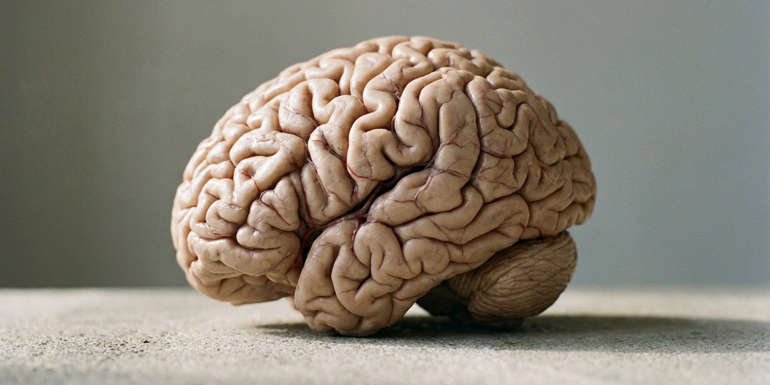
In young men with autism spectrum disorders, the physical arrangement of brain folds in areas associated with social and emotional processing appears to be altered. Recent analyzes of brain imaging data show that neurotypical boys often exhibit skewed folding patterns that are less common in boys with autism. The researchers published their research in a journal cerebral cortex.
The human brain is characterized by a wrinkled outer layer known as the cerebral cortex. This structure is filled with raised ridges and deep grooves. Together, these folds work to maximize the amount of neural tissue that can fit within the narrow spaces of the human skull.
Valleys or grooves pushed deep into the brain tissue are called sulci. Most of the surface area of the cerebral cortex is actually buried within these hidden grooves. Because the cortex regulates higher mental functions, scientists are actively studying the shape and location of these folds to better understand human cognition and neural development.
One particular subregion of this outer layer is the anterior cingulate cortex. This region serves as a central hub for emotional regulation, cognitive control, and social cognition. These represent broad areas of mental processing that are often affected in individuals with autism spectrum disorder (ASD).
A prominent anatomical feature extending within the anterior cingulate cortex is the paracingulate sulcus. The paracingulate sulcus is a tertiary brain fold that runs parallel to the major sulcus in that region. Unlike some major brain folds that appear virtually identical in all humans, this particular sulcus shows extreme physical differences across populations.
Some people develop long, prominent paracingulate sulci in both the left and right hemispheres of the brain. Some are completely missing this secondary fold on both sides. If there are folds, their exact shape and trajectory vary widely from person to person.
In neurotypical populations, the presence of the paracingulate sulcus is usually asymmetric. People frequently develop this fold in the left hemisphere of the brain, while the right hemisphere remains relatively smooth in that particular area. Previous research has shown that this shift in left-to-right patterns correlates with performance on executive function tasks and the ability to guess what others are thinking.
The researchers wanted to map the paracingulate sulcus in autistic people because these cognitive traits closely overlap with different manifestations of autism. Neuroscientists Ethan Willebrand and Enrique Martinez from the University of Wisconsin-Madison and the University of California, Berkeley led the research team. They aimed to outline the exact characteristics of this particular groove in young people with autism.
The researchers used existing structural magnetic resonance imaging (MRI) scans of 200 young men between the ages of 5 and 18. Half of the participants had previously been diagnosed with an autism spectrum disorder. The other half were neurotypical.
To ensure that the analytical framework was robust and accurately represented reality, the scientists divided these participants evenly into a primary discovery group and a secondary replication group. This split-sample method allows researchers to test initial statistical models on completely separate batches of data.
A trained rater manually evaluated each participant’s MRI scan to determine the presence or absence of the paracingulate sulcus in both hemispheres. Folds must be at least 20 millimeters long and 4 millimeters deep to be officially classified as functionally present. Manually defining the limits of elusive folds is recognized as the gold standard in neuroanatomical research.
In addition to checking for the basic presence of folds, the team used computer algorithms to extract precise geometric proportions. They measured the total length of the paracingulate sulcus by following its longest unbroken path. They also calculated the maximum depth of the sulcus and the average thickness of the gray matter lining the sulcus.
This analysis revealed consistent differences in how the paracingulate sulcus is distributed between the left and right hemispheres. Neurotypical participants are likely to have an asymmetric folding pattern, typically with sulci on the left side of the brain but no sulci on the right side. In contrast, participants with autism spectrum disorder showed increased structural symmetry.
Specific left-handed asymmetry was significantly reduced in participants with autism. They were much more likely to have a matching set of features, either having grooves on both sides of the brain or uniformly lacking grooves on both sides. The odds of having an asymmetric paracingulate sulcus were significantly higher in typically developing boys than in autistic boys.
This structural difference remained constant even when researchers adjusted their statistical models to account for potential confounding variables. The team controlled for participants’ age, measured intelligence quotient, and physical location of the medical center where the MRI scan was performed.
Although the overall structural symmetry behaved differently between groups, the specific physical dimensions of the grooves did not. Statistical tests showed that the length, depth, and cortical thickness of the paracingulate sulcus did not differ between autistic and neurotypical brains. These specific geometric measurements were not statistically significant in either the primary discovery group or the replication group.
This contrast highlights the well-known distinction between different features of human neuroanatomy. Tertiary brain folds, such as the paracingulate sulcus, begin to form internally well before birth, usually around the 36th week of human gestation. This structural blueprint reflects the very early biological constraints placed on the developing fetal brain.
Such early formation suggests a prenatal origin of the symmetry differences observed in autistic youth. The relatively symmetrical arrangement seen in autistic brains may indicate early biological mutations in genetic factors or cellular machinery that determine how the fetal brain physically folds. Once these basic folds are established in utero, their layout remains largely stable throughout life.
On the other hand, measurements such as fold depth and outer gray matter thickness are highly dynamic. Cortical thickness changes throughout childhood development, shrinking or growing in response to life experiences, learning, and physical maturation. These dynamic measures did not differ between groups, leading the researchers to suggest that neuroanatomical differences associated with autism operate primarily at a distance, rooted in a person’s prenatal development.
Although anatomical variation is noteworthy, the current study has multiple limitations. Participants included only young men under 20 years of age. There is enormous biological diversity in autism spectrum disorders, and brain folding patterns are sometimes known to differ significantly based on biological sex. This means that the researchers’ findings cannot simply be generalized to women or older adults with autism.
Furthermore, the researchers were unable to directly link anatomical differences in this precise population to specific behavioral or cognitive traits. The public image database they relied on did not contain details of a uniform cognitive test for all 200 participants. A dedicated follow-up project is needed to understand how this central brain fold symmetry actually influences everyday mental tasks.
Manually mapping the folds of the human brain also takes considerable time. This limits the total number of scans that scientists can reasonably analyze in a single anatomical project. The researchers recommend that future research invest directly in developing automated computer-based tools that can precisely track folds in the brain that are not instinctively and universally present.
These advanced technologies allow anatomical experts to process thousands of scans simultaneously. This will ultimately help map the highly variable physical landscape of the human brain on a much larger scale, revealing exactly how tiny prenatal folds shape behavior throughout a person’s lifespan.
The study, “Anterior cingulate diffraction patterns are altered in autism spectrum disorders,” was authored by Ethan H. Willbrand, Enrique Martinez, Jacob J. Ludwig, Samira A. Maboudian, and Kevin S. Weiner.
