


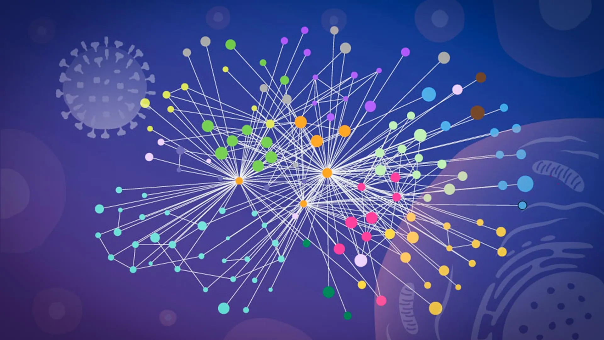
Researchers at EMBL Hamburg, in collaboration with scientists at the Leibniz Institute for Molecular Pharmacology (FMP), have created an unusually detailed map showing how influenza A reshapes infected human cells. Their customized workflow allowed them to directly observe protein interactions within intact cells, rather than relying solely on disaggregated samples.
Seasonal influenza causes 3 million to 5 million severe cases and 650,000 deaths worldwide each year. Influenza A has also caused multiple pandemics, including the 1918 Spanish flu pandemic.
After a virus enters a cell, it releases RNA that contains instructions for making a small set of viral proteins. These proteins spread into the host cell, reorienting its molecular systems and turning the cell into a site for producing new virus particles.
Mapping influenza virus within intact cells
A clearer picture of the acquisition could help scientists develop more effective influenza vaccines and antiviral drugs. To achieve that goal, researchers need to know which viral proteins interact with human proteins, where those contacts occur, and how the virus uses them to support its own replication.
This new study is the first to map direct contacts between influenza proteins and human proteins on a large scale within intact, infected cells. The structural details were also precise enough to allow researchers to model how the interacting proteins bind.
“Our study provides a new way to study influenza-host interactions in their native context with structural insights,” said Jan Kosinski, group leader at EMBL Hamburg and the Center for Structural Systems Biology (CSSB). “The current results are a snapshot of moments during infection and open the door to studying influenza-host interactions throughout the infection cycle.”
Overcoming major experimental problems
Tracking protein-protein interactions during active infection is extremely difficult. Much of the previous work relied on biochemical techniques, which required scientists to break up cells before measuring protein contacts.
The process can distort what was happening inside living cells. Once the internal compartment is disrupted, proteins that were originally isolated can come into contact in the lab. At the same time, weak, transient, or location-specific interactions may disappear. As a result, researchers may have a hard time determining which connections were actually present during the infection.
“This is when we learned that our collaborators Boris Bogdanou and Huang Liu from FMP Berlin had developed a specialized version of cross-linking mass spectrometry (XL-MS), a long-established technique for mapping protein contacts, specifically tailored to virus-infected cells,” Kosinski said.
A special method gave the team the breakthrough it needed. This made it possible to capture interactions that occur only briefly within specific regions of infected cells.
“XL-MS allows us to capture protein-protein interactions directly in infected, intact cells, while also providing structural information about how these interactions occur,” explained Bogdanow, currently a junior research group leader at the Charité Institute for Virology at the University of Berlin. “This provides insight into the interface between viruses and human cells and, through structural modeling, may help identify viable targets for future pharmaceutical intervention.”
Combining experimental data with AlphaFold
The researchers combined the XL-MS results with computational structural modeling. This allowed them to identify the interacting viral and human proteins and estimate how those proteins are arranged when they bind.
To build these structural models, the research team used a modified version of AlphaFold, a Nobel Prize-winning protein structure prediction algorithm.
“The main advantage of the modified AlphaFold approach is that experimental cross-linking data can be directly fed into structural modeling,” Kosinski explained. “This tells the model which parts of the virus and host proteins are in close proximity to each other in infected cells. This was particularly useful for virus-host complexes, which are often difficult to predict reliably.”
Two ways influenza hijacks human cells
The survey results are natural microbiologyrevealed two notable strategies that influenza A appears to use when controlling cells.
The first focuses on hemagglutinin, a protein on the surface of the virus. Influenza uses hemagglutinin to attach to and invade host cells. The researchers tracked the protein’s passage through the cell’s internal transport and processing networks.
This network is made up of compartments that fold, modify, and prepare proteins before sending them to their final location. The analysis showed that several human proteins help properly fold and modify hemagglutinin during infection. Some of these host proteins have previously had poorly understood functions.
Influenza dissolves structures within the nucleus
The second discovery concerned paraspeckles, small droplet-like compartments located inside the cell nucleus. The research team found that influenza A infection caused these structures to dissolve.
When the paraspeckles disintegrated, the RNA-binding proteins held inside were released. The virus may then use those proteins to support its own replication.
“What surprised us the most was the paraspeckles,” said Julia Kotova, a former predoctoral researcher in the Kosinski group at EMBL Hamburg, now at ETH Zurich, and lead author of the paper. “When we observed that these small organelles in the nucleus lysed consistently in every cell line and every influenza strain we tested, we knew this might be a strategy rather than a side effect of infection.”
This disruption could have multiple benefits for influenza.
“Viruses may also have a second benefit. There is also evidence to suggest that paraspeckles contribute to cellular stress responses and the regulation of antiviral genes, so destroying paraspeckles may also weaken some of the cell’s defense responses,” Kosinski added.
Initiatives through collaboration between three institutions
The project relied on technology and expertise shared by the three institutions. The researchers carried out the cross-linking mass spectrometry work at the Charité in Berlin. Glycoproteomic analyzes were completed at the EMBL Proteomics Core Facility.
The team performed AlphaFold modeling on the EMBL Compute Cluster, and microscopy imaging was performed at CSSB’s Advanced Light and Fluorescence Microscopy (ALFM) facility.
A new way to study potential pandemic viruses
The findings show that studying the molecular contacts inside intact, infected cells can reveal how and where the virus controls human cellular machinery. Researchers say this kind of “mapping in context” could also help explain how other viruses behave.
“Although the exact host factors and mechanisms often differ from virus to virus, we believe our overall approach of mapping native virus-host interactions at specific stages of infection by combining intracellular cross-linking, structural modeling, and follow-up of target cell biology remains broadly applicable,” Kosinski said.
Although the study looked at lab-adapted strains of influenza, the researchers believe the same strategy could eventually be used to investigate viruses with greater pandemic potential.
Professor Bogdanow agrees. “Although this study focuses on laboratory-adapted strains, this study lays the groundwork for applying this methodology to potentially pandemic-relevant viruses such as H5N1 and uncovering the interaction networks that support virus replication within human cells.”
