


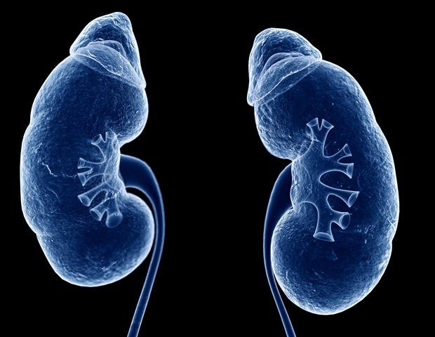
A new case report has been published in Volume 17. onco target July 2, 2026, titled “Renal Oncocytoma: Alpha Case Report and Literature Review.”
The study was led by first author and corresponding author Areti Kalftsou of the Second Department of Internal Medicine, Aticon General Hospital, National Kapodistrian University of Athens, Greece.
Renal oncocytoma is a rare benign tumor of the kidney that accounts for approximately 5-9% of renal epithelial tumors. Although it is considered benign, it closely resembles kidney cancer on imaging tests, making accurate diagnosis difficult. Because treatment strategies differ significantly between benign oncocytomas and malignant renal tumors, establishing the correct diagnosis is essential to avoid unnecessary surgery while ensuring appropriate patient care.
In this report, researchers describe the case of an 82-year-old woman who had a 2-month history of gross hematuria. Contrast-enhanced computed tomography (CT) identified a 55 × 34 mm exophytic lesion arising from the upper pole of the left kidney. Based on the imaging features, this mass was initially classified as a Bosniak category IV renal lesion, raising a strong suspicion of renal malignancy.
Instead of proceeding directly to surgery, the clinical team performed a CT-guided percutaneous biopsy. Histopathological examination, supported by immunohistochemical analysis, established the diagnosis of renal oncocytoma. The tumor showed positive staining for E-cadherin and cyclin D1, focal positivity for cytokeratin 7 (CK7), and negative staining for CD117, carbonic anhydrase IX (CAIX), vimentin, and RCC, findings supporting the final diagnosis.
After multidisciplinary discussion, the tumor board recommended percutaneous cryoablation due to the relatively small size and slowly progressive nature of the lesion. This minimally invasive approach was particularly appropriate given the patient’s clinical situation. More than 2 years after treatment, annual imaging shows no evidence of disease progression.
The authors also discuss the wide range of diagnostic challenges associated with renal oncocytoma. Conventional CT and magnetic resonance imaging can localize these tumors but often cannot reliably differentiate them from chromophobe renal cell carcinoma or other renal tumors. While histopathological evaluation remains the gold standard for diagnosis, percutaneous biopsy is increasingly recognized as a safe and minimally invasive method to establish a diagnosis before deciding on treatment.
This report also discusses advances in pathological diagnosis. Immunohistochemical markers such as CK7 and cyclin D1, together with characteristic histological features, can help distinguish renal oncocytoma from chromophobe renal cell carcinoma and oncocytic/chromophobe hybrid tumors and improve diagnostic confidence in difficult cases.
”This case supports an individualized treatment strategy for renal oncocytoma, balancing tumor characteristics, patient comorbidities, and potential treatment risks to achieve optimal results.. ”
According to the authors, management decisions must be made on an individual basis, taking into account tumor size, patient age, symptoms, and overall health. For certain elderly patients and those at high surgical risk, minimally invasive approaches such as percutaneous cryoablation may enable effective disease management while preserving renal function and reducing treatment-related morbidity.
Overall, this case highlights the importance of combining imaging, biopsy, histopathology, immunohistochemistry, and multidisciplinary evaluation when managing renal masses. The findings demonstrate how accurate diagnosis can guide personalized treatment decisions and help some patients achieve superior clinical outcomes while avoiding unnecessary surgery.
sauce:
Reference magazines:
Kalfzo A. Others. (2026) Renal oncocytoma: A case report and literature review. Onco target. DOI: 10.18632/oncotarget.28893. https://www.oncotarget.com/article/28893/text/
