


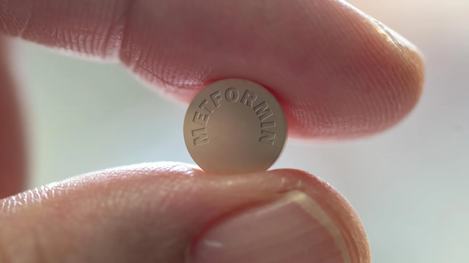
Metformin has been the standard treatment for type 2 diabetes for more than 60 years, but scientists did not fully understand how metformin lowers blood sugar levels. Now, researchers at Baylor College of Medicine and collaborators around the world have identified an unexpected factor behind that effect: the brain. Their findings reveal a previously unknown brain pathway involved in metformin’s antidiabetic effects, opening the door to more precise and effective treatments. This study scientific progress.
“It is widely accepted that metformin lowers blood sugar levels primarily by reducing glucose excretion in the liver. Other studies have shown that metformin acts through the intestine,” said corresponding author Dr. Makoto Fukuda, associate professor of pediatric nutrition at Baylor University. “We investigated the brain because it is widely recognized as an important regulator of systemic glucose metabolism. We investigated whether and how the brain contributes to metformin’s antidiabetic effects.”
The brain’s role in blood sugar control
The researchers focused on a protein called Rap1, located in a region of the brain known as the ventromedial hypothalamus (VMH). They found that metformin’s ability to lower blood glucose levels at clinically relevant doses was dependent on suppression of Rap1 activity in this region.
To investigate this further, the research team used genetically engineered mice lacking Rap1 in the VMH. These mice were fed a high-fat diet as a model for type 2 diabetes. Low doses of metformin did not improve blood sugar levels. In contrast, other antidiabetic drugs such as insulin and GLP-1 agonists remain effective, suggesting a specific role for Rap1 in metformin’s actions.
Big effect with just a small amount of brain administration
The scientists then tested whether the brain itself could cause these effects. They injected tiny doses of metformin directly into the brains of diabetic mice. This treatment significantly lowered blood sugar levels, even at doses thousands of times lower than oral intake.
“We also investigated which cells in the VMH are involved in mediating the effects of metformin,” said Professor Fukuda. “We found that SF1 neurons were activated when metformin was introduced into the brain, suggesting that SF1 neurons are directly involved in the drug’s effects.”
How metformin activates brain cells
The researchers measured the electrical activity of these neurons using brain tissue samples. Metformin increased activity in most of these cells, but only in the presence of Rap1. In mice lacking Rap1 in these neurons, the drug had no effect. This showed that Rap1 was required for metformin to activate these brain cells and lower blood sugar levels.
“This discovery has changed the way we think about metformin,” said Professor Fukuda. “It’s not just acting on the liver and intestines, it’s also acting on the brain. It takes high concentrations of the drug for the liver and intestines to respond, but we found that the brain responds at much lower levels.”
New directions in diabetes treatment
Only a few diabetes drugs are known to affect the brain, but this study suggests that metformin has been working for some time. The findings present new opportunities to develop treatments that directly target this brain pathway.
“These findings open the door to the development of new diabetes treatments that directly target this pathway in the brain,” Professor Fukuda said. “Additionally, metformin is also known for other health benefits, such as slowing brain aging. We plan to investigate whether this same brain Rap1 signaling is also involved in the drug’s other well-documented effects on the brain.”
Research contributors and funders
Other contributors to this work include Hsiao-Yun Lin, Wesheng Lu, Yanlin He, Yukiko Fu, Kentaro Kaneko, Peimen Huang, Ana B De la Puente-Gomez, Chunmei Wang, Yongjie Yang, Feng Li, and Yong Xu. The authors are affiliated with one or more of the following institutions: Baylor College of Medicine, Louisiana State University, Nagoya University (Japan), and Meiji University (Japan).
This study was supported by the National Institutes of Health (R01DK136627, R01DK121970, R01DK093587, R01DK101379, P30-DK079638, R01DK104901, R01DK126655), USDA/ARS (6250-51000-055), American Supported by a grant from Heart. Association (14BGIA20460080, 15POST22500012) and American Diabetes Association (1-17-PDF-138). Additional support was provided by the Uehara Memorial Foundation, the Takeda Science Foundation, the Foundation for Applied Enzymology, and the NMR and Drug Metabolism Core at Baylor College of Medicine.
