


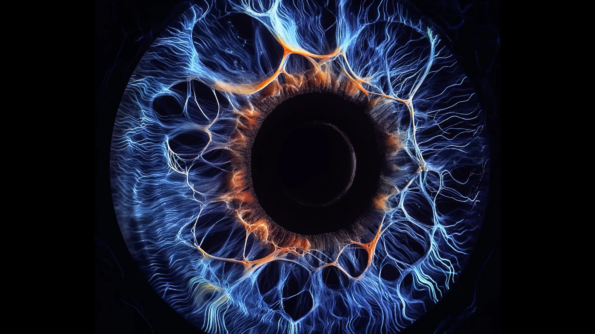
New research from Yale School of Medicine (YSM) suggests that the eye processes visual information in a much more intimate way than scientists thought. The discovery challenges long-held ideas about how visual signals travel across the retina and may help explain how we detect faint objects and see in the dark.
Our visual system quickly analyzes various features of a scene, such as color, contrast, movement, and shape. This process, known as parallel visual processing, allows the brain to interpret complex images almost instantly by sending different types of information along separate paths.
Researchers have long believed that these pathways remain largely independent as visual signals travel through the retina to the brain. However, new research published in 2016 shows that neuronfound that these channels are closely linked through hidden electrical connections. According to the researchers, this cooperation could strengthen weak visual signals before they travel deeper into the visual system.
“We found that while different channels can provide unique functionality, they are also interconnected by underlying electrical circuits,” says Dr. Yao Xue, a postdoctoral fellow in YSM’s Department of Ophthalmology and Visual Sciences and lead author of the study.
Bipolar cells form an unexpected communication network
Vision begins when the rods and cones of the retina detect light. These specialized cells pass information to neurons known as bipolar cells. At this stage, visual information is sorted into a dozen parallel channels that process features such as daylight, night vision, color, contrast, and shape.
When researchers took a closer look at synapses, the tiny junctions where bipolar cells communicate, they discovered something unexpected. Instead of remaining isolated, channels that were supposed to be separate were sharing information with each other.
Neurons communicate through two main types of synapses: chemical synapses and electrical synapses. Chemical synapses use neurotransmitters to transmit messages between cells, while electrical synapses, also called gap junctions, transmit signals through direct current. Bipolar cells were generally thought to rely primarily on chemical communication.
A new study finds that electrical synapses connect most of these separate information channels in both mouse and human retinas. When the researchers electrically stimulated a single bipolar cell, the response spread far beyond that one pathway. Rather than neurotransmitter release being restricted to one channel, they observed a cloud-like, widespread pattern of activity, revealing widespread communication between different bipolar cell types.
“When we stimulated one bipolar cell, many bipolar cells released neurotransmitters,” said lead researcher Z. Jimmy Chou, Ph.D., Marvin L. Sears Professor of Ophthalmology and Visual Sciences.
The researchers also identified one type of bipolar cell, known as BC6, that appears to play a leading role in coordinating this network. Signals originating from BC6 spread through multiple visual pathways in an organized and hierarchical pattern.
“People used to think that different types of bipolar cells were more or less autonomous,” Zhou says. “But we discovered drivers within all of these cell types that form this hierarchical network.”
Scientists say this combination of specialized pathways and electrical communication provides the retina with the best of both approaches. Individual channels allow you to focus on specific visual features, and channels can be connected to share information when the signal is particularly weak.
“If the signal is already very weak and split into multiple channels, there is very little that can be processed in each channel,” says Seunghoon Lee, Ph.D., a researcher in YSM’s Department of Ophthalmology and Visual Sciences and co-author of the study. “This integration is particularly useful for detecting low-contrast signals or signals from very small objects.”
“And cells don’t randomly cooperate,” Xue added. “Among them is a commander – BC6 – who is directing them to relay the signal to downstream targets.”
Record signals on the intact retina
To map these communication networks, the team combined several experimental techniques. They used advanced imaging to monitor how bipolar cells responded by releasing neurotransmitters, while simultaneously stimulating individual cells and recording the responses of neighboring cells.
Bipolar cells have long been difficult to study because they reside deep in the retina. Previous experiments required slicing the retina to reach it, a process that could disrupt the natural circuitry the researchers wanted to examine.
In this study, the Yale team successfully used a dual patch clamp technique on a completely intact mouse retina. The researchers used electrodes to stimulate specific bipolar cell types and simultaneously recorded how neighboring cells responded.
“No other laboratory in the world has been able to do this kind of recording systematically,” Zhou says. “This is a masterpiece of Yao Xue’s doctoral thesis research, combining an innovative approach with outstanding electrophysiological skills.”
The researchers then repeated the experiment using intact human retina obtained through the Department of Pathology’s Legacy Tissue Donation Program. The researchers say these are the first experiments of their kind to be performed on intact human retinas.
What the discovery means
Because the retina is part of the central nervous system, researchers say these findings may have implications beyond vision. Understanding how retinal circuits process information may provide new insights into how other neural networks in the brain function.
The research could also advance scientists’ understanding of diseases that damage the retina, such as macular degeneration, glaucoma, and congenital night blindness.
The researchers also say the study highlights the value of curiosity-driven science. Rather than testing a single predefined idea, this experiment uncovered a previously unknown mechanism that changes the way scientists think about visual processing.
“While our experiments did not start with a specific hypothesis, we uncovered a fundamental processing mechanism in the visual system,” says Lee. “This is an important reminder of how important curiosity-driven research is to discovery.”
