

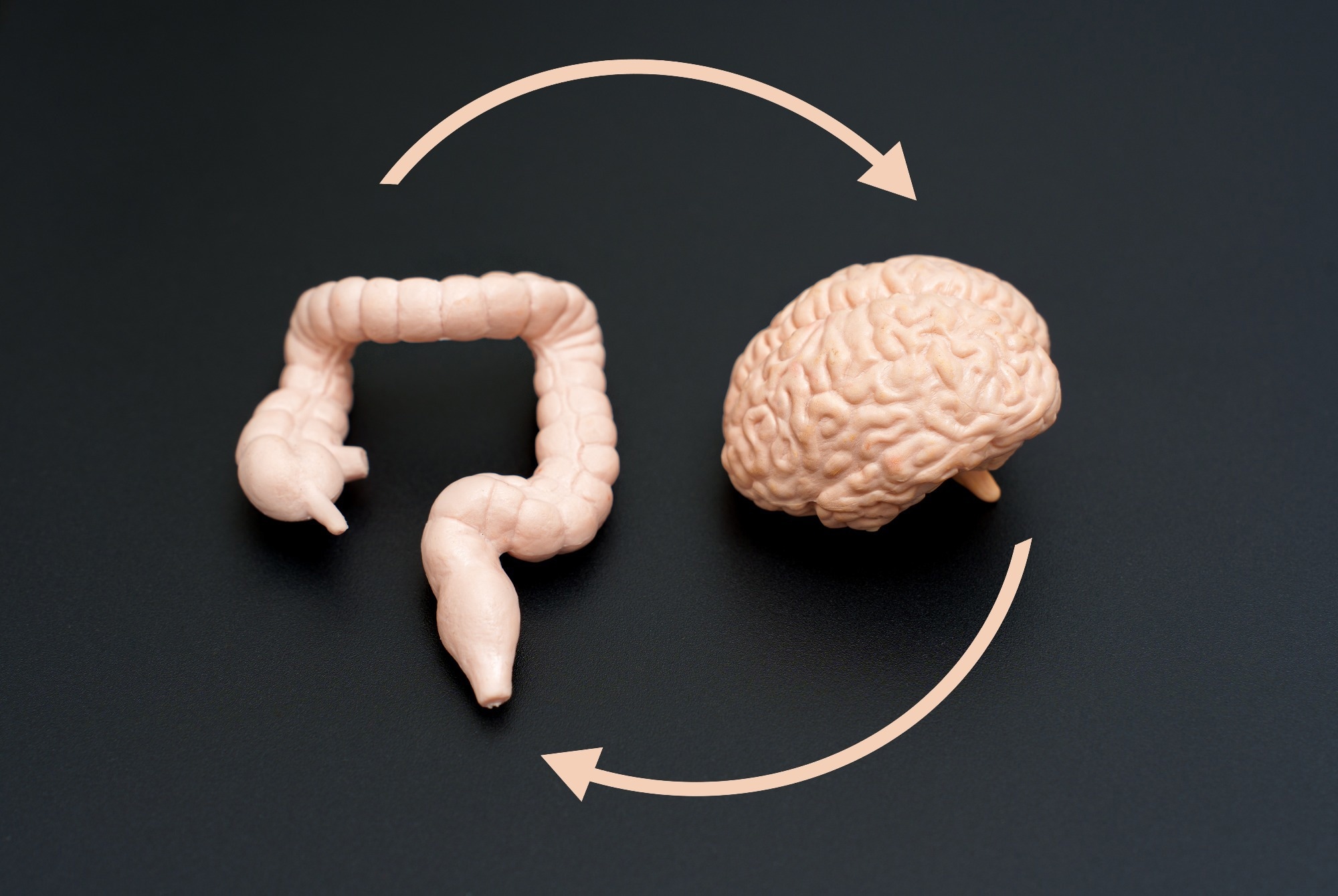
A new study in Nature shows how inflammation caused by parasites in the gut activates the tuft cell-to-serotonin-to-vagus nerve pathway, helping to explain why infections suppress appetite.
Research: Parasites cause epithelial cell crosstalk and promote gut-brain signaling. Image credit: Chizhevskaya Ekaterina / Shutterstock
In a recent study published in the journal naturea group of researchers investigated how intestinal epithelial cells interact to cause changes in gut-brain signaling and food intake during parasite infection.
Did you know that intestinal infections can affect how much you eat and even how you feel? The gastrointestinal tract acts as a sensory system that detects harmful stimuli and communicates with the brain.
Specialized epithelial cells such as enterochromaffin (EC) cells and tuft cells are important for sensing irritants and parasites. EC cells release serotonin (5-HT), which activates neural pathways associated with pain and nausea. Tuft cells, on the other hand, detect parasites and mount an immune response. However, it is still unclear how these cells work together to influence brain signal transmission.
Further research is needed to explain how the gut-brain connection influences feeding-related responses during infection.
Tuft cell and EC cell research design
In this study, we combined cellular, molecular, and animal-based experimental approaches to examine the communication between intestinal tuft cells and EC cells. The structure and function of the intestinal epithelium were replicated using mouse intestinal tissue organoids.
Calcium imaging technology was applied to monitor cell activation using genetically encoded indicators such as genetically encoded calcium indicator (GCaMP) and serotonin sensors such as genetically encoded GPCR activation-based 5-HT sensor (gGRAB5-HT).
To assess acetylcholine release during experiments, we generated biosensor cells expressing various receptors, including muscarinic acetylcholine receptor (mAChR) subtype 1 (M1R) and 5-hydroxytryptamine receptor 3 (5-HT3). We then studied them using electrophysiological methods such as patch-clamp techniques to assess the electrophysiological properties of tuft cells and demonstrated that tuft cells do not exhibit conventional forms of excitability. Several pharmacological agents were used to isolate the functional muscarinic and nicotinic receptor pathways involved in acetylcholine release from biosensor cells.
Animal models included genetically modified mice that lack tuft cells or lack choline acetyltransferase, the enzyme needed to make acetylcholine. Type 2 immune responses were induced with interleukin-25 and parasitic infections were modeled using: Nippostrongillus brasiliensis.
The researchers recorded neural activity in vagal afferent fibers in vitro from enteric nerve preparations. Food intake and other spontaneous behaviors were measured to assess the physiological effects of gut-brain signaling.
Acetylcholine in tuft cells activates serotonin release
This discovery reveals a previously unrecognized communication pathway between tuft cells and EC cells that links immune responses to neural signaling. Tuft cells were shown to release acetylcholine through two different mechanisms. Tuft cells were shown to release acetylcholine through two different mechanisms. First, they rapidly released acetylcholine through TRPM5-dependent signaling in response to the protist-derived signal succinate. Subsequently, a sustained “leak-like” release of acetylcholine was demonstrated during type 2 inflammation induced by interleukin-4 or interleukin-25.
Tuft cells can release acetylcholine without synaptic vesicles or electrical excitability. This means that tuft cells have their own special way of releasing neurotransmitters. Instead of activating all mAChRs, acetylcholine released from tuft cells only activates muscarinic receptors on crypt EC cells, primarily mAChR subtype 3 (M3R). Activation of M3R generates intracellular calcium, which subsequently stimulates serotonin release.
It was observed that the magnitude and duration of acetylcholine release controlled the effect. Acute acetylcholine release produced limited serotonin output, insufficient to strongly activate vagal nerve fibers. In contrast, sustained release of acetylcholine during inflammation increased serotonin levels and strongly activated vagal afferent neurons via 5-HT3 receptors.
This signaling pathway was experimentally demonstrated in genetically modified mice that depend on tuft cells and acetylcholine synthesis. Mice lacking tuft cells or choline acetyltransferase showed decreased serotonin release and reduced neural activation. Additionally, pharmacological inhibition of muscarinic receptors reduced stimulation of EC cells, highlighting the importance of cholinergic stimulation.
Reduced food intake due to parasitic infection
Nippostrongillus brasiliensis Parasitic infections further corroborated these findings in the physiological environment. This includes increased serotonin levels in the intestinal crypts, enhanced vagal activity, and activation of receptors in brainstem regions including the nucleus of the solitary tract. This pathway was absent or severely attenuated in mice with tuft cell dysfunction or acetylcholine synthesis deficiency.
Sustained activation of the gut-brain connection can reduce food intake. Although the effects of acute stimulation of tuft cells were minimal, type 2 inflammation reduced food intake, particularly during the peak period of inflammation rather than immediately after infection. This suggests that the intestine uses persistent signaling to transmit ongoing infections to the brain, triggering adaptive responses that may help limit the nutrients available to the parasite.
Relationship between the gut-brain axis and feeding
This study shows that intestinal tuft cells and EC cells form an important communication network linking immune detection of parasites and brain-mediated changes in feeding behavior. When tuft cell-derived acetylcholine activates muscarinic receptors on crypt EC cells, this pathway promotes serotonin release, which stimulates vagal afferent neurons and influences feeding behavior during infection. The release of acetylcholine in two distinct time phases supports the idea that the gastrointestinal tract has mechanisms to distinguish between short-term and long-term threats.
These findings provide new insights into the gut-brain axis and have important implications for understanding changes in feeding behavior and gastrointestinal disease processes, as well as neuroimmune interactions. Therapeutic strategies targeting identified pathways may ultimately provide innovative approaches to treat symptoms and metabolic disorders associated with infectious diseases.
Reference magazines:
- Touhara, KK, Xu, J., Castro, J., Liang, HE, Li, G., Brizuela, M., Harrington, AM, Garcia-Caraballo, S., Neumann, D., Rossen, ND, Deng, F., Schober, G., Li, Y., Locksley, RM, Brierley, SM, and Julius, D. (2026). Parasites cause epithelial cell crosstalk and promote gut-brain signaling. Nature, 1-9. DOI: 10.1038/s41586-026-10281-5, https://www.nature.com/articles/s41586-026-10281-5