


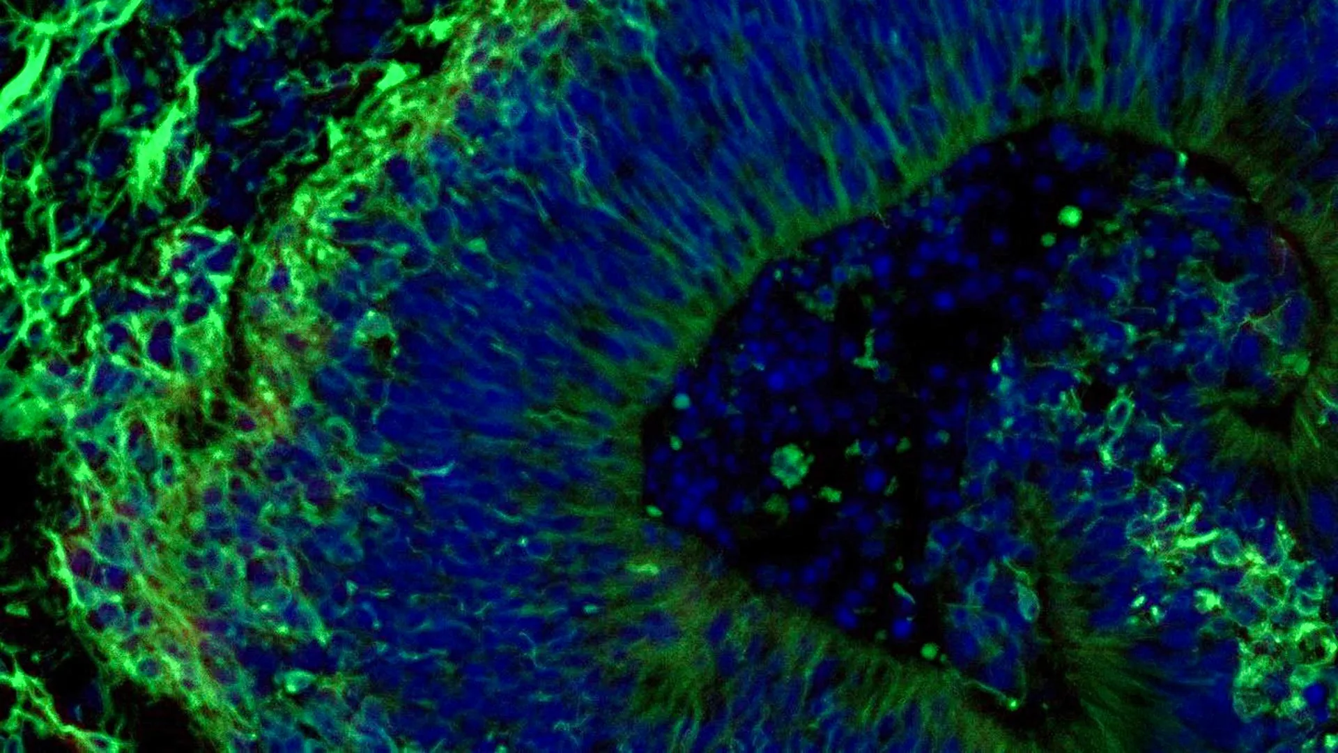
Scientists at the Johns Hopkins School of Medicine have discovered new evidence that small chunks of brain tissue grown from the cells of Alzheimer’s disease patients could help researchers predict how different patients will respond to medications used to manage psychiatric symptoms associated with Alzheimer’s disease.
The study focused on lab-grown brain tissue called organoids. The discovery adds to the evidence that these miniature brain models may ultimately help scientists develop and select more precise treatments for specific groups of Alzheimer’s patients. Alzheimer’s disease is the most common dementia, affecting more than 7 million Americans.
The researchers also discovered that the organoids release tiny particles called extracellular vesicles that carry cellular information. These particles may provide new biomarkers for diagnosing Alzheimer’s disease and determining how advanced the disease is.
This research was partially funded by the National Institutes of Health; Alzheimer’s Disease and Dementia: Journal of the Alzheimer’s Association.
Mini-brain models could support personalized care
“Our study suggests that large-scale patient-derived brain organoids and the vesicles they secrete may be useful in staging Alzheimer’s disease, investigating the mechanisms that cause it, and assessing how subgroups of patients respond to different treatments,” said study leader Dr. Vassiliki Machairaki, associate professor of medical genetics at the Johns Hopkins University School of Medicine.
There is currently no cure for Alzheimer’s disease. However, selective serotonin reuptake inhibitors (SSRIs) are often prescribed to help manage neuropsychiatric symptoms such as anxiety, depression, and agitation. Although these symptoms affect nearly all patients, responses to medications vary widely, Macieraki says.
Researchers at Johns Hopkins studied miniature models of the hindbrain. The hindbrain is an area at the back of the skull that helps control important functions such as breathing, sleep, and heart rate. The research team wanted to determine whether these models could reveal molecular signs that indicate whether the SSRI escitalopram oxalate can help reduce symptoms associated with Alzheimer’s disease.
Converting a patient’s blood cells into brain tissue
The researchers started with blood samples taken with permission from Alzheimer’s patients at the NIH-funded Johns Hopkins Alzheimer’s Disease Research Center.
They reprogrammed blood cells back to a stem cell-like state. These cells are called induced pluripotent stem cells and can grow into any type of cell in the body.
Using induced pluripotent stem cells from Alzheimer’s patients and healthy people, the researchers created hindbrain organoids containing specialized brain cells, or neurons, that produce the neurotransmitter serotonin.
The cells were induced to organize into small pea-sized clusters of brain tissue that resembled the hindbrain. The study included hundreds of organoids representing individual patients with Alzheimer’s disease and healthy participants. Professor Machairaki believes this may be one of the largest brain organoid studies ever carried out in Alzheimer’s disease research.
Alzheimer’s disease organoids show distinct molecular changes
Patient-derived organoids recapitulated several key biological features of Alzheimer’s disease at the molecular level.
Compared to organoids made from healthy people, organoids grown from cells from Alzheimer’s patients showed differences in proteins involved in communication between brain cells, inflammation, and pathways associated with Alzheimer’s disease.
The researchers then treated the organoids with escitalopram oxalate, a widely prescribed antidepressant.
In some patient-derived organoids, the drug increased proteins involved in serotonin signaling and transmission between brain cells. These are the pathways that antidepressants are designed to affect. Other organoids showed little or no molecular reaction.
“We used these organoids to model how the tissues of some patients respond to commonly prescribed SSRIs,” Macieraki says. “At a large-scale level, our model could ultimately be used to identify subgroups of patients who are more likely to respond to certain drugs based on the underlying molecular mechanisms, which in the long term could aid in the development of precise targeted therapies.”
Tiny vesicles may reveal drug reactions
The research team then investigated whether the extracellular vesicles released by the organoids could serve as biomarkers for Alzheimer’s disease or help researchers assess a tissue’s response to treatment.
The scientists examined proteins in extracellular vesicles released from patient-derived organoids and healthy control organoids before and after treating the organoids with escitalopram.
The vesicles contained proteins involved in essential brain activities, such as communication between neurons, memory, and the release of neurotransmitters.
Organoids grown from cells from Alzheimer’s patients showed clear changes in several disease-related proteins. Levels of RAB3A, NSF, and ATCAY were lower in Alzheimer’s disease organoids. These proteins play an important role in normal signaling between brain cells.
Levels of some proteins increased in certain samples after escitalopram treatment. This change was particularly pronounced in proteins associated with serotonin signaling and synaptic pathways that are targeted by antidepressants.
Some organoids showed strong molecular responses, while others showed little or no change. Machairaki says this change raises the possibility that extracellular vesicles in brain organoids could eventually help identify patients most likely to benefit from certain treatments.
Building more realistic brain organoids
Professor Machairaki plans to develop more sophisticated organoids containing immune cells and blood vessel-like networks that mimic blood vessels. Adding these features could make the tissue more similar to living human brain tissue.
With further research, she hopes that extracellular vesicles may one day function as a type of liquid biopsy. Such tests can help diagnose Alzheimer’s disease, determine the stage, and identify a patient’s specific disease subtype.
Professor Machairaki emphasized that this study is an early step towards that goal.
In addition to Machairaki, scientists contributing to the study include Rachel Boyd, Daiyun Dong, Ram Sagar, Waqar Ahmed, Zenia Androni, Paul Rosenberg, Konstantin Riquesos, and Kenneth Witwer of Johns Hopkins University, Anton Ilyuk of Timora Analytical Operations, and Anton Polsteinsson of the University of Rochester School of Medicine and Dentistry.
Funding for this research was provided by the National Institutes of Health (T32 AG058527, R01AG052510, P30AG066507, 1RF1AG083801, AGR01054771, AGR01050515, AGR01046543, AGR01071522), Paul G. Allen Frontiers Foundation, and the Richman Family Precision Medicine Center of Excellence Alzheimer’s Disease Research Center at Johns Hopkins University.
In accordance with Johns Hopkins University policy, none of the authors declares any relevant conflicts of interest.
