


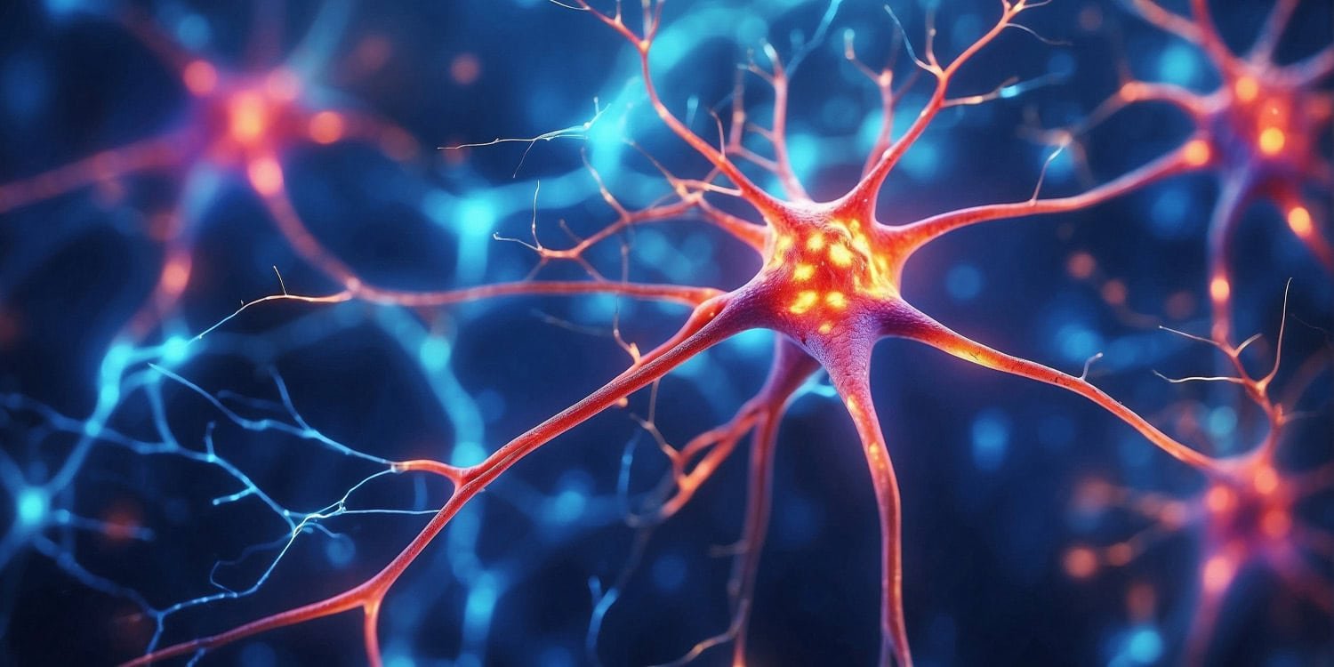
Recent research published in Proceedings of the National Academy of Sciences The results suggest that individual cells in the human brain have computational capabilities far greater than those found in other mammals. By applying artificial intelligence to model these brain cells, scientists have discovered that human neurons are highly sophisticated information processing units in their own right. These findings provide evidence that humans’ unique cognitive abilities may derive from the complex structure and function of individual cells, not just the vast number of cells within brain networks.
The brain is made up of billions of individual cells called neurons, which communicate with each other to process information. Most of humans’ advanced cognitive functions, such as language and problem solving, occur in the cerebral cortex. This is the wrinkled outer layer of the brain. Within the cortex, the primary cells responsible for transmitting excitatory signals are called pyramidal neurons. These cells are named for their characteristic cone-shaped cell bodies.
Neurons receive incoming electrical signals through branch-like structures called dendrites. The signal travels through the dendrites to the cell body. When the combined signals reach a certain threshold, the neuron fires an electrical pulse known as a spike or action potential, which transmits the message to other cells. The way neurons integrate these input signals and decide whether to fire or not is essentially a type of microscopic computation. These tiny cellular decisions form the biological basis of all human thought and behavior.
Previous anatomical observations have shown that human cortical pyramidal neurons look physically different from rodent neurons. Human neurons tend to be larger and have more extensive and intricately branched dendrites. But scientists lacked a standardized way to accurately measure how these physical differences affect a cell’s ability to process information.
A research team led by scientists from the Hebrew University of Jerusalem and Vrije Universiteit Amsterdam aimed to measure the functional complexity of these microscopic brain cells. They sought to determine whether the unique physical characteristics of human neurons actually lead to higher computational power compared to rat neurons. To do this, they needed a tool that could quantify how well a neuron can convert multiple input signals into a single output spike.
To solve this problem, scientists have developed a new metric called the functional complexity index. This index relies on machine learning concepts. Machine learning is often used to find patterns in large consumer datasets, but here it is used as a yardstick to measure biological complexity. The central idea is to train a standard artificial neural network to mimic the behavior of biological brain cells. Artificial neural networks are computer systems designed to recognize patterns, loosely inspired by the structure of the brain.
The researchers reasoned that if biological neurons functioned like simple switches, small-scale artificial networks could easily learn how to predict their behavior. When biological neurons perform highly complex calculations, the same artificial networks have a hard time replicating their output. The lower the performance by the artificial network, the higher the functional complexity index score of biological cells.
Scientists conducted experiments using detailed digital models of biological neurons. They utilized three-dimensional reconstructions of 24 specific cells. This sample contained 12 human cortical pyramidal neurons and 12 rat cortical pyramidal neurons. The selected cells represented different depths of the brain cortex, specifically spanning layers 2, 3, 4, 5, and 6.
The researchers generated a huge dataset for each of the 24 digital neurons. They ran a simulation in which they exposed a digital cell to a random input electrical signal spread across the dendritic branches. Each simulation lasted 10 seconds, and 12,000 simulations were performed for each neuron. This generated data equivalent to more than one day of continuous neural activity data for each individual cell model.
The researchers then built a standard artificial neural network featuring three internal processing layers, each containing 128 computational units. They fed this network with exactly the same received signal used in the simulation. The artificial network was then tasked with predicting the precise millisecond timing of electrical spikes generated by the biological cell model.
The authors found that human cortical neurons had significantly higher complexity index scores than rat cortical neurons. Artificial networks had much more difficulty predicting the timing of spikes in human cells. This suggests that human neurons perform much more complex transformations of input signals than rat neurons.
To understand the cause of this difference, the research team analyzed 58 separate physical measurements of the cells’ dendritic branches. They found that total dendrite surface area was the single strongest predictor of cell complexity score. The length of branches branching into other branches was also a major factor. This provides evidence that larger, more expansive dendritic structures allow different parts of the cell to process information somewhat independently, greatly increasing overall computational power.
The researchers also investigated the role of synapses, the tiny connection points where signals enter dendrites. Specifically, we focused on the NMDA receptor. These are specialized proteins located at synapses that respond nonlinearly to incoming electrical signals. This means that when enough signals arrive at once, NMDA receptors dramatically amplify the current, rather than simply adding the signals together.
The researchers tested both rat-like and human-like synaptic properties in digital simulations. Scientific evidence suggests that human excitatory synapses contain more NMDA receptors and respond more sharply to voltage changes. When researchers applied these human-like synaptic properties to their models, they significantly increased the complexity of the cells’ functions. The combination of sprawling dendrites and highly responsive NMDA receptors tends to push the processing power of human neurons to much higher levels.
The data also revealed interesting changes in how complexity is distributed across cortical layers. In the rat model, neurons located in layer 5 were the most complex. In the human model, neurons in layers 2 and 3 were significantly more complex than neurons in other layers. Layers 2 and 3 are known to be particularly expanded in the human brain, suggesting an evolutionary adaptation in the way the human brain allocates its computational resources.
Although this study details single-cell calculations, there are several limitations. The study relied entirely on computer simulations of neurons, rather than living tissue in an active brain. Due to the current lack of experimental data on the specific electrical properties of human dendrites, the model does not include all possible active ion channels found in living cells. This means that digital cells may behave slightly differently than biological cells in a real human brain.
Furthermore, the functional complexity index is highly dependent on the specific design of the artificial neural network used for testing. If the artificial network is too shallow or deep, the difference in scores between cells can be compressed. The researchers chose a three-tier network as a middle ground, but the exact numbers may vary for different computer architectures.
Future research directions could include investigating other anatomical features, such as small projections on dendrites known as spines, to see how signal processing changes. The researchers also hope to apply this new measurement tool to other types of brain cells and to different species, such as non-human primates. Ultimately, collecting data from living human brain cells in a laboratory setting could help scientists validate the computational patterns observed in these digital simulations.
The study, “Dendrite morphology and synaptic nonlinearity enhance the functional complexity of human cortical neurons,” was authored by Ido Aizenbud, Daniela Yoeli, David Beniaguev, Christiaan PJ de Kock, Michael London, and Idan Segev.
