


Despite the known effects of smoking on other microbial communities, this study suggests that the ocular surface ecosystem may remain surprisingly stable, leaving the possibility of small changes that can be detected in larger studies.
Study: Effects of smoking on the human ocular surface microbiome and tear proteome. Image credit: komokvm / Shutterstock
In a recent study published in the journal scientific reportResearchers from the University of Bern in Switzerland assessed whether smoking is associated with changes in the ocular surface microbiome and tear proteome by comparing microbial composition, diversity, functional profiles, and tear proteins between smokers and non-smokers.
background
Did you know that although smoking can affect the microbial community in your body, its effects on your eyes are largely unknown?
The ocular surface microbiome is a low-biomass community of bacteria, viruses, fungi, and other eukaryotes that may help support ocular health by modulating local immune responses, maintaining epithelial barrier integrity, and limiting pathogen colonization. When this balance is disrupted, it can lead to symptoms such as dry eye disease, conjunctivitis, and keratitis.
Although smoking is known to be an important risk factor for the development of various eye diseases, the specific effects of smoking on the ocular surface microbiome have not been systematically investigated.
About research
The researchers recruited 41 adults, including 17 smokers and 24 non-smokers, from the Department of Ophthalmology at Bern University Hospital in Switzerland. Smokers had smoked at least six cigarettes daily for at least 2 years, whereas nonsmokers had no history of tobacco use recorded.
After obtaining written informed consent, samples were collected in winter, spring, and summer. Tears were collected using the Schirmer type I tear test, and pooled conjunctival swabs were taken from both eyes for microbiome analysis. Positive and negative controls were included to ensure data quality.
Microbial DNA extracted from conjunctival swabs was removed from human sequences and contaminants and then subjected to shotgun metagenomic sequencing to identify bacterial, fungal, and viral communities. Microbial functional analyzes including gene family and pathway profiling were also performed.
Tear fluid samples were analyzed by nanoliquid chromatography-tandem mass spectrometry to determine their proteomic profiles. Statistical analyzes included alpha and beta diversity assessment, principal coordinate analysis (PCoA), permutation multivariate analysis of variance (PERMANOVA), differential abundance analysis, principal component analysis (PCA), and differential expression analysis with correction for multiple testing. A post hoc power analysis was also conducted to estimate the ability to detect significant differences between study groups.
Research results
A total of 41 conjunctival swab samples were sequenced, including 24 from nonsmokers and 17 from smokers. No significant differences were observed between groups in age or gender.
After decontamination with the microDecon pipeline, the main bacterial groups in non-smokers were Actinobacteria (59.1%), Proteobacteria (25.8%), and Firmicutes (13.9%), and Proteobacteria (43.2%), Actinobacteria (35.6%), and Firmicutes (20.7%) were the main bacterial phyla in non-smokers. Smoker.
In general, Cutibacterium acnes was relatively more prevalent in non-smokers (52.1%) and smokers (30.7%), while Moraxella osloensis accounted for 20.9% and 30.6%, respectively. Limosilactobacillus fermentum and Sphingobium yanoikuyae were also frequently detected.
Among eukaryotes, both Basidiomycota and Ascomycota were major groups. In particular, Basidiomycota accounted for 51.0% of the eukaryotic presence in non-smokers and approximately 64.1% in smokers, and Ascomycota accounted for 49.0% and 35.9%, respectively.
Saccharomyces cerevisiae was the most common eukaryotic species, accounting for 44.9% and 27.3%, respectively, and Malassezia globosa was the second most common species, accounting for 25.8% and 27.7%, respectively.
Furthermore, Cryptococcus neoformans contributed to 18.0% and 23.1% of the eukaryotic abundance in both groups. Finally, there was some variation within the virus community. Since phylum-level annotations were not available in the virus dataset, the researchers examined the viral community at the order level.
The most commonly occurring viral orders included unclassified viruses, Caudovirales, and herpesviruses. Results also revealed sequences assigned to Glypta fumiferanae ichnovirus (21.3% in non-smokers and 14.3% in smokers), Ictaluride herpesvirus type 1 (11.1%) in both groups, and BeAn 58058 virus (8.7% in non-smokers and 15.8% in smokers).
Bacterial diversity analysis showed no significant differences between smoker and non-smoker groups. The mean bacterial Shannon diversity index was 0.89 in non-smokers and 0.81 in smokers (p = 0.5235).
Eukaryotic alpha diversity index showed no significant difference between the two study groups (p = 0.369), while viral alpha diversity remained similar between groups (p = 0.83). PCoA results showed that bacterial, eukaryotic, and viral communities were not clearly clustered. PERMANOVA also showed no significant differences for bacteria (R2 = 0.052; p = 0.106), eukaryotes (R2 = 0.034; p = 0.488), or viruses (R2 = 0.035; p = 0.175).
Differential abundance analysis did not identify bacterial, eukaryotic, or viral taxa that were significantly different after correction for multiple testing. However, one gene-level trait of bacteria has been reported to differ between groups, suggesting that the lack of significant taxonomic differences should not be extended to all functional microbial traits.
Post hoc power analysis showed that the power observed across bacterial, fungal, and viral alpha diversity comparisons was consistently low. The authors therefore cautioned that this finding should not be interpreted as proof of bioequivalence between smokers and non-smokers, but rather as evidence against large smoking-related effects. Additional analyzes also showed that sampling time, DNA extraction kit, and pollen allergy status explained more of the variation in bacterial communities than smoking behavior.
Proteomics analysis of tear fluid identified 1,066 proteins, of which 1,065 were quantified using iMaxLFQ with a missing value rate of 0.1% before filtering across 40 samples. The median coefficient of variation of the studies was found to be 8.7%, while the mean coefficient of variation of the studies was 8.0%. PCA results showed no significant separation between samples obtained from smokers and non-smokers (PERMANOVA p = 0.116, R2 = 0.035).
Furthermore, differential expression analysis showed that no statistically significant proteins remained after correction for multiple comparisons. Because we used conservative thresholds in our proteomic analysis, small smoking-related changes in tear protein abundance could not be excluded.
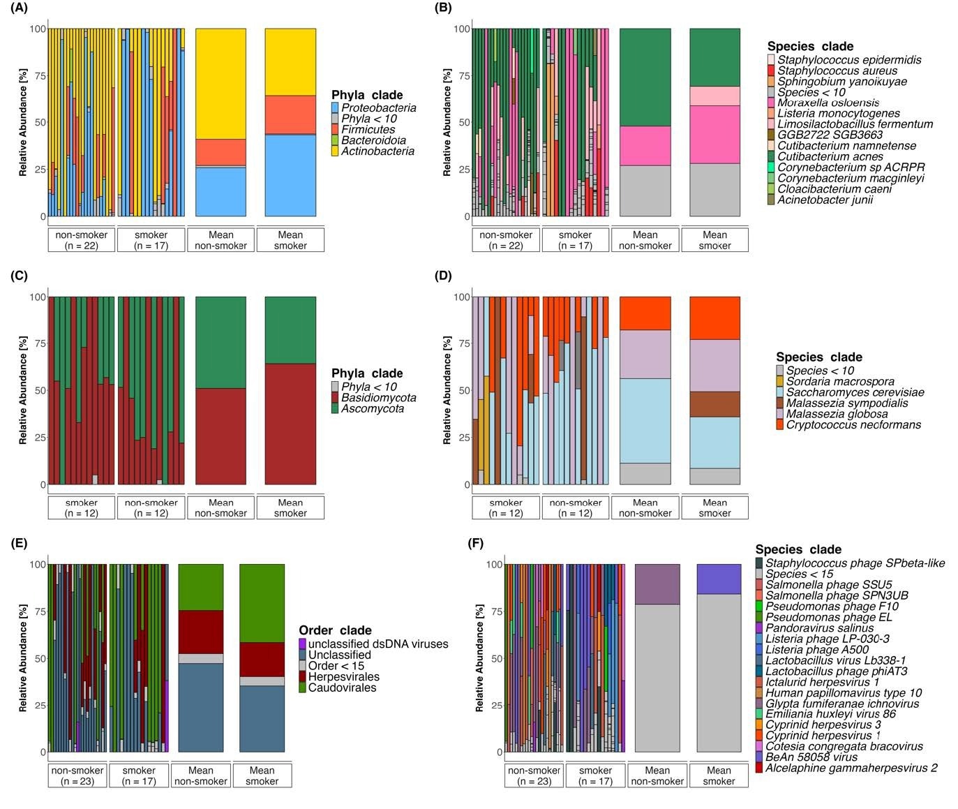
(A-F) Taxonomic profiles of the ocular surface microbiome in non-smokers (bacteria n = 22, eukaryotes n = 12, viruses n = 23) and smokers (bacteria n = 17, eukaryotes n = 12, viruses n = 17). Bar plots show the relative abundances of bacterial (AB), eukaryotic (CD), and viral (EF) taxa at the phylum level (left) and species level (right). Individual bars represent samples. Two additional bars indicate the group average. Colors indicate different taxa.
conclusion
Results showed that in this small cohort, smoking was not associated with significant or consistent changes in taxonomic composition, microbial diversity, or ocular surface tear proteome. After correction for multiple testing, neither bacterial, eukaryotic, or viral taxa nor tear protein markers showed significant differences between the two populations. The tear proteome remains largely stable, consistent with resilient host-microbe interactions on the ocular surface.
The authors concluded that the ocular surface microbiome appears to maintain ecological stability despite smoking, but stressed that this result does not exclude smaller effects related to smoking. Future longitudinal studies with larger sample sizes are needed to better understand environmental and disease-related factors that may influence ocular surface microbial balance. Further research is needed to see how daily habits affect ocular surface health.
Reference magazines:
- Federico AO Silva Gutierrez, SC Morandi, N. Eldridge, MS Zinkelnagel, DC Gissetbri. (2026). Effects of smoking on the human ocular surface microbiome and tear proteome. scientific report. Doi: 10.1038/s41598-026-60743-z, https://www.nature.com/articles/s41598-026-60743-z
